General Information about Tamoxifen
Tamoxifen is a drugs that has been used for decades within the treatment of breast cancer. It is commonly prescribed for girls who've been diagnosed with breast most cancers that has unfold to other elements of the physique. Tamoxifen is a sort of hormonal therapy that works by blocking the consequences of estrogen within the body. This hormone is thought to promote the growth of certain types of breast cancer, so by blocking its results, tamoxifen can decelerate and even stop the expansion of cancer cells.
While tamoxifen has proven to be useful in the remedy of breast most cancers, it does come with its own risks and side effects. The most common unwanted effects include scorching flashes, fatigue, and vaginal dryness. More critical side effects can include blood clots and an increased threat of uterine cancer. It is essential for patients to discuss their medical history with their physician earlier than starting tamoxifen therapy to make certain that it is safe for them.
In conclusion, tamoxifen is a crucial treatment in the battle in opposition to breast most cancers. It is a hormonal therapy that works by blocking the effects of estrogen within the body, stopping the growth of cancer cells. It has been proven to scale back the danger of breast cancer recurrence and may also be used to treat different conditions. However, like any medicine, it does include dangers and unwanted side effects, and should solely be taken underneath the steerage of a health care provider. With continued research and developments in the area of breast cancer therapy, tamoxifen stays an integral part of the fight towards this disease.
Aside from its use in treating breast most cancers, tamoxifen has also been found to be efficient for different conditions. For example, it can be used to treat gynecomastia, a situation in which males develop breast tissue because of hormonal imbalances. Tamoxifen works by blocking estrogen receptors within the breast tissue, lowering the dimensions of the breast and enhancing symptoms.
In addition, tamoxifen can interact with other medications, so it is important for patients to inform their physician of some other medicines they're taking. This includes over-the-counter drugs, dietary supplements, and herbal cures. Patients must also keep away from taking tamoxifen throughout being pregnant, as it could hurt the creating fetus.
One of the principle advantages of tamoxifen is that it could reduce the danger of breast most cancers recurrence. Studies have proven that ladies who take tamoxifen for 5-10 years after their initial remedy have a 50% decrease threat of their most cancers coming back. This is due to the ability of tamoxifen to forestall estrogen from fueling the growth of most cancers cells. In addition, tamoxifen can also shrink existing tumors, making them simpler to remove throughout surgical procedure.
Tamoxifen is often prescribed after surgery and other remedies, corresponding to chemotherapy, have been accomplished. It is also used for girls who're at an elevated threat of developing breast most cancers, both as a result of family historical past or other danger components. This medicine just isn't efficient in ladies who've estrogen-receptor-negative breast cancer, as it solely works by blocking the results of estrogen.
To harness the ability of transcription factors to reprogram cells in a clinically relevant manner menstruation 4 weeks postpartum buy tamoxifen 20 mg low cost, Ieda et al. They showed that direct reprogramming of fibroblasts resulted in the stable shift of the epigenetic signature of several cardiac genes-Actn2, Ryr2, and Tnnt2. Heart Muscle Development Successful development of the cardiovascular system requires a complex integration of multiple inputs that ultimately results in stable gene expression profiles that determine the identity of the differentiated cell. Transcription factors interact with epigenetic factors to define how developmental cues can be translated into stable and heritable cardiac gene expression profiles. Brg1 is necessary for early cardiomyocyte cell proliferation as well as the differentiation into mature cardiac myocytes. Murine models with cardiac-specific deletion of Brg1 have a thinned myocardium and absent interventricular septums. In addition to regulating cell cycle progression, Brg1 is also necessary for the differentiation of cardiac myocytes. Thus, Brg1 is important in the normal progression of differentiation from fetal to adult cardiac myocytes. These studies suggest that the effective development of cardiac myocytes requires an interaction between transcription factors and epigenetic complexes. The inappropriate expression of Six1 resulted in massive right ventricular hypertrophy and mild pulmonary stenosis in adult mice. Basic science and human clinical trials have demonstrated that a transient exposure to hyperglycemia results in a higher incidence of diabetic vascular disease despite a return to normal glucose levels. Because epigenetic mechanisms can induce stable changes in gene expression that persist despite the subsequent absence of the inciting stimulus, investigators have explored epigenetic mechanisms to explain the legacy effect. The increase in p65 results in the persistent expression of a proinflammatory gene expression profile. Next, using human heart failure samples, they confirmed that human heart failure is also associated with a significant shift in genome-wide H3K4me3 and H3K9me3 profiles. They found that the activating H3K4me3 marks are associated with many important canonical signaling pathways. Taken together, these results suggest that there is an association between H3K4me3 marks and H3K9me3 marks and the alteration in gene expression profiles that accompanies cardiac disease states. To investigate the mechanistic role of histone-modifying proteins in the development of cardiac hypertrophy, Zhang et al. H3K27 methylation marks are associated with transcriptionally repressed chromatin. These mice demonstrated lethal congenital heart malformations including compact myocardial hypoplasia, hypertrabeculation, and ventricular septal defects. They also showed that cardiacspecific overexpression of Jmjd2a resulted in an exaggerated hypertrophic response. Human studies reveal that Brg1 is upregulated in cardiac tissue from patients with hypertrophic cardiomyopathy. The degree of Brg1 expression correlates with the extent of the hypertrophic phenotype. Brg1 is normally downregulated in adult heart tissue; however, pressure overloadinduced hypertrophy in murine models results in re-expression of Brg1. This work demonstrated that it is necessary to maintain epigenetic profiles in fully differentiated tissues, and that an inability to maintain these marks over time can be a mechanistic cause of disease. Conclusion Epigenetic phenomenon in cardiac development and disease are currently under intense investigation because epigenetic phenomena provide a mechanistic explanation for how environmental factors can cause changes in the cardiac phenotype. As outlined earlier, considerable evidence supports the important role of epigenetic mechanism in the developing heart. These same epigenetic mechanisms that establish stable and heritable gene expression profiles during development also seem to be important contributors to the development of disease. Epigenetic mechanisms may be particularly important in the development of adult cardiac disease states because most cardiac myocytes are post-mitotic terminally differentiated cells. Therefore, an inability to maintain the epigenome over the life of a cell may alter gene expression profiles in aged myocytes. These shifts in the epigenome could predispose to or initiate the development of disease. In addition, epigenetic mechanisms may provide a rational explanation for why some disease processes appear reversible and some are irreversible. In any case, a deeper understanding of epigenetic mechanisms will provide us with new insights into cardiac biology. Ringrose L, Paro R: Polycomb/trithorax response elements and epigenetic memory of cell identity. Czermin B, Melfi R, McCabe D, et al: Drosophila enhancer of zeste/esc complexes have a histone h3 methyltransferase activity that marks chromosomal polycomb sites. Beisel C, Imhof A, Greene J, et al: Histone methylation by the drosophila epigenetic transcriptional regulator ash1. Nakamura T, Mori T, Tada S, et al: All-1 is a histone methyltransferase that assembles a supercomplex of proteins involved in transcriptional regulation. Takahashi K, Yamanaka S: Induction of pluripotent stem cells from mouse embryonic and adult fibroblast cultures by defined factors. Yazawa M, Hsueh B, Jia X, et al: Using induced pluripotent stem cells to investigate cardiac phenotypes in timothy syndrome. Delgado-Olguin P, Huang Y, Li X, et al: Epigenetic repression of cardiac progenitor gene expression by ezh2 is required for postnatal cardiac homeostasis. He A, Ma Q, Cao J, et al: Polycomb repressive complex 2 regulates normal development of the mouse heart. Retinopathy and nephropathy in patients with type 1 diabetes four years after a trial of intensive therapy. The diabetes control and complications trial/epidemiology of diabetes interventions and complications research group.
After injection orbital compression for 10 to 15 minutes is applied with superpinky or any other method pregnancy years after vasectomy discount 20 mg tamoxifen otc. The anaesthetic solution used for peribulbar anaesthesia consists of a mixture of 2% lignocaine, and 0. During general anaesthesia for ocular surgery, use of relaxants, endotracheal intubation and controlled respiration is preferred. Lid speculums non co-operative and mentally retarded adults, perforating ocular injuries, major operations like. It retracts the lids and its solid blades keep the lashes away from the field of operation. Uses: It is used to hold the superior rectus muscle while passing a bridle suture under it; to stabilize the eyeball during any operation such as cataract surgery, glaucoma surgery, corneal surgery, etc. Eye speculums are used to keep the lids apart during: · Intraocular operations such as cataract surgery, glaucoma surgery and keratoplasty. Use: these are used to hold the cornea or scleral edge (of incision) for suturing during cataract, glaucoma, repair of corneal and/or scleral tears and keratoplasty operations. Use: these are used to catch the iris for the purpose of iridectomy during operations for cataract, glaucoma, optical iridectomy and excision for iris prolapse, tumours and entangled foreign bodies. It is applied near the limbus to hold the conjunctiva and episcleral tissue together. Uses: these are used to epilate the cilia in trichiasis and stye, to remove cilia after electrolysis and cryolysis and to remove cilia lodged in the punctum. It has multiple straight grooves (at right angle to the limbs) near the tip and a locking mechanism near the ringed end. The small-sized artery forceps, also called as mosquito artery forceps are more commonly used in ophthalmology. Uses: (i) To catch the bleeding vessels during operations of the lids and lacrimal sac. Use: It is used to retract the lids during examination of the eyeball in cases of blepharospasm in children, in cases with marked swelling and ecchymosis, removal of corneoscleral sutures, removal of corneal foreign body and for double eversion of upper lid to examine the superior fornix. It is made up of two limbs with three curved pins on each for engaging the edges of the skin incision. It consists of a handle with a curved blade (which conforms to the pupillary margin) at one end. Use: It is used to take measurements during squint, ptosis, retinal detachment and pars plana vitrectomy surgery. These are available in various sizes with straight or curved tips, in different shapes and may be with or without locking system. The jaws of the needle holder are finely serrated to hold the fine needles firmly. Use: Spring type needle holders are used for passing sutures in the conjunctiva, cornea, sclera and extraocular muscles. It is a long, narrow, thin and straight blade with a sharp tip and cutting edge on one side. Uses: (i) Previously, it was used for making an abinterno corneoscleral incision during cataract surgery and for iridectomy operation. These are large needle holders and all are of similar type with slight model differences. Use: these are very commonly used in lid surgery and also for passing superior rectus suture. Use: Keratomes are used to make valvular corneal incisions for entry into the anterior chamber for all modern techniques of cataract extraction viz. It is a divider- shaped needle with sharp cutting edges resembling in appearance to a small keratome. It has a short flat blade with a semicircular blunt dissecting edge, which is bevelled on both the surfaces like a chisel. Uses: (i) It can be used to separate the conjunctiva and sub conjunctival tissue from the sclera and limbus when limbal based flap is made for trabeculectomy surgery. Presently, presterilized razor blade fragments mounted on a disposable plastic handle. Use: It is the most commonly used cutting device for making incisions in cataract, glaucoma, keratoplasty, sclerotomy, pterygium and many other operations. It is used to make a small valvular clear corneal incision (commonly called as side port incision) in phacoemulsification and other intraocular surgeries including pars plana vitrectomy. It is a fine straight but triangular knife similar to 15° side port entry blade but with cutting edges on both sides. Its blade is curved and either mounted on a plastic handle (disposable) or can be fixed with metallic handle. Presently, disposable cystitome is prepared by bending the disposable 26 gauge or 30 gauge hypodermic needle. Use: It is used for doing anterior capsulotomy or capsulorhexis during extracapsular cataract extraction. Use: It is used to cut and undermine conjunctiva in various operations and to undermine skin during operations on lids and lacrimal sac. Use: It is used to perform iridectomy, iridotomy and to cut the prolapsed form vitreous and pupillary membrane. Use: They are used as a handy alternative to plain straight and plain curved ringed scissors for cutting and undermining conjunctiva in various operations and to cut sutures. Uses: (i) these are used to enlarge corneal or corneoscleral incision for conventional intra capsular and extra capsular cataract extraction techniques (sparingly performed procedures nowadays) of cataract surgery. Uses: (i) these are used for cutting anterior capsule of the lens in extra capsular surgery and for cutting 100 nylon sutures. They are large, stout and strong scissors having curved sharp blades with blunt ends.
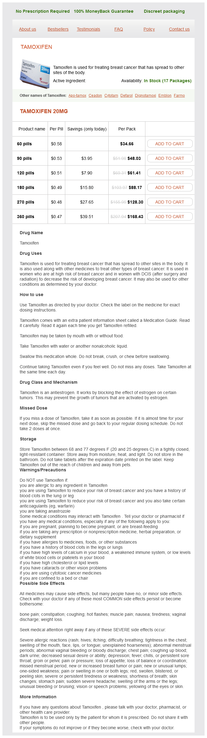
Tamoxifen Dosage and Price
Tamoxifen 20mg
- 60 pills - $34.66
- 90 pills - $48.03
- 120 pills - $61.41
- 180 pills - $88.17
- 270 pills - $128.30
- 360 pills - $168.43
The anterior leaflet is then taken down menopause sleep buy tamoxifen now, in particular, capitalizing on the true area of delamination between the anterior leaflet and the right ventricle. All fibrous and muscular attachments of the leading edge of the leaflet are extensively taken down, with attention paid not to divide true chordal support to the leading edge. The dissection is then extended clockwise to include the septal leaflet all the way to the anteroseptal commissure; the septal leaflet often is quite diminutive (nearly absent) and may have multiple fenestrations that require closure. With the anterior, inferior, and septal leaflets all fully mobilized, the cut edge of the inferior leaflet is rotated clockwise to the proximal edge of the septal leaflet (as in the Carpentier repair), and the two are approximated with interrupted 6-0 prolene sutures completing the "cone". In doing so, there are now 360 degrees of leaflet tissue that can comprise the new tricuspid P. With the cone retracted, the area of atrialized right ventricle is closed vertically, with attention paid to the preservation of the right coronary artery (which lies just outside this area). As with the Carpentier repair, vertical plication (unlike horizontal plication) will preserve the height of the true tricuspid annulus, and not artificially displace it further downward. Note the location of the "true" tricuspid annulus to which the repaired valve will be secured, as well as the vein of D, which often denotes the region of the conduction system. Attention needs to be paid to the depth of these sutures so as not to compromise the right coronary artery. Interrupted sutures are placed carefully to eliminate the blind pouch of the excluded atrialized segment. The newly constructed "cone" (tricuspid valve) is then reattached at the level of the true annulus; here several interrupted sutures are recommended as reinforcement should a P. Often the annulus must be plicated to meet the smaller circumference of the "cone. Additional support of the Cone Not infrequently, additional plication sutures-or if the patient is of adult size, an entire annuloplasty ring-are required to fully support the cone. Any residual leak or leaflet fenestrations are addressed, as is the patent formen ovale (shown). In the schematic form, the reconstructed cone is now at the level of the true annulus, the atrialized right ventricle having been vertically plicated maintains this height, and the annulus itself is downsized and supported. Need for right ventricular unloading To note, if the right ventricle appears inadequate to support full cardiac output, a Glenn cavopulmonary connection can be created to offload some of this volume. Tricuspid Valve Replacement When the abnormality produces obstruction within the right ventricle, the tricuspid valve is excised and replaced with an appropriate prosthesis. If more than mild to moderate tricuspid insufficiency is present following valve repair, replacement is indicated. The septal and posterior leaflet tissues are resected, but the anterior leaflet tissue is often incorporated in the suture technique of anchoring the prosthesis. Because of the ambiguous location of the conduction system owing to displacement of the tricuspid valve, the true atrial wall above the coronary sinus is used to construct a new annulus to which the prosthesis is sutured with multiple, interrupted, and everting mattress sutures of 2-0 Tevdek buttressed with pledgets (see Chapter 8). Alternatively, a patch of glutaraldehyde-treated autologous or bovine pericardium may be sewn to the right atrial wall, beginning at the anteroseptal commissure, continuing above the level of the atrioventricular node and inside the coronary sinus, back to the posterior annulus. The valve sewing ring is then attached to this patch to avoid placing sutures in the septal annulus. A ventricular septal defect is often present and may be associated with left ventricular outflow obstruction due to malalignment of the conal septum. Other associated anomalies may include a bicuspid aortic valve, truncus arteriosus, and aortopulmonary window. The interruption may be just distal to the left subclavian artery (type A), between the left carotid and left subclavian arteries (type B), or between the innominate and left carotid artery (type C). Type B is the most common form of interrupted aortic arch, and type C is very rare. Hypoplasia of the proximal arch between the innominate and left carotid arteries is defined as a diameter less than 60% of that of the ascending aorta. The distal arch between the left carotid and left subclavian arteries is considered hypoplastic if the diameter is less than 50% that of the ascending aorta. A hypoplastic aortic arch may be associated with a ventricular septal defect and other congenital heart lesions. Patients with an interrupted or hypoplastic aortic arch usually present as neonates when the ductus arteriosus closes and flow to the descending aorta ceases or is severely restricted. Infusion of prostaglandin E1 is immediately started to reopen the ductus arteriosus to perfuse the distal aorta. One-stage complete repair of the aortic arch and associated cardiac defects is the preferred technique. Most of the thymus gland (if present) is removed to allow adequate mobilization of the branches of the aortic arch. Cannulation Traditionally, deep hypothermic arrest has been used for surgery involving the aortic arch. More recently, lowflow antegrade cerebral perfusion has been advocated during reconstruction of the arch to avoid or minimize circulatory arrest and cerebral ischemia. A purse-string suture is placed on the far right side of the distal ascending aorta near the origin of the innominate artery. In patients with an interrupted aortic arch, a second purse-string suture is placed on the proximal main pulmonary artery. The right and left pulmonary arteries are dissected and encircled with Silastic tourniquets. Dual arterial cannulation with flexible 8 to 10-French aortic cannulas is achieved using a Y-connector on the arterial line for interrupted aortic arch. The right atrial cannula is then placed through a purse-string suture on the right atrial appendage. As cardiopulmonary bypass is initiated, the snares on the right and left pulmonary arteries are tightened to prevent flooding of the pulmonary bed.