General Information about Procardia
Procardia has been proven to be efficient within the treatment of angina. In a research carried out by the University of California, Procardia was found to significantly lower the frequency and severity of angina attacks. It was additionally shown to improve exercise tolerance and increase blood circulate to the center. Additionally, Procardia has been discovered to be as efficient as different generally prescribed drugs for angina, similar to beta-blockers and calcium channel blockers.
In conclusion, Procardia is a generally prescribed medicine for the therapy of angina. It has been confirmed to effectively scale back the frequency and severity of angina attacks and enhance train tolerance. However, as with every medicine, there could additionally be potential side effects and precautions that have to be taken. It is necessary to consult a well being care provider before beginning Procardia and to intently observe dosage directions. Procardia, when used appropriately, can be a extremely effective treatment for angina, offering relief and enhancing the quality of life for individuals who suffer from this condition.
Precautions:
As with any medication, there could also be unwanted facet effects associated with utilizing Procardia. The most typical unwanted effects are delicate and embody dizziness, headache, flushing, and nausea. These signs are often short-term and should subside as the physique adjusts to the medication. More critical unwanted effects, although uncommon, may include low blood strain, irregular heartbeat, and swelling of the ankles or feet. In some circumstances, Procardia can also worsen pre-existing conditions, corresponding to coronary heart failure or liver disease. It is necessary to consult a well being care provider if any regarding or persistent unwanted aspect effects happen.
Before taking Procardia, it may be very important inform your physician of any pre-existing medical conditions, allergy symptoms, and medications you're at present taking. Procardia could interact with sure medicine, including beta-blockers, digoxin, and some antibiotics. It can be necessary to avoid consuming grapefruit or grapefruit juice whereas taking Procardia, as it might improve the amount of medication in your bloodstream and trigger undesirable side effects. Procardia just isn't really helpful to be used during pregnancy or breastfeeding.
Procardia, also referred to as nifedipine, is a prescription medication generally used for the treatment of angina. Angina is a sort of chest ache that occurs when the guts does not receive enough oxygen-rich blood. This pain can be extreme and is usually described as a tightness, pressure, or squeezing sensation within the chest. Procardia works by stress-free the blood vessels, allowing more blood and oxygen to flow to the center. This article will talk about the makes use of, effectiveness, unwanted facet effects, and precautions of utilizing Procardia for angina.
Procardia is primarily used for the treatment of angina. It is efficient in relieving chest ache brought on by coronary artery illness, a medical condition where the arteries that offer blood and oxygen to the heart turn out to be slender or blocked. Procardia helps to prevent angina assaults by relaxing the blood vessels, which reduces the workload of the guts and will increase blood move to the guts. It may additionally be used to deal with high blood pressure, also identified as hypertension.
Side Effects:
Uses:
Effectiveness:
Involved individuals may have no problems except in the presence of some special stress blood vessels under skin discount procardia online visa, such as infection or surgery. Patients with no symptoms at all for a period of years can suddenly develop coma, convulsions, and death following an apparently mild infection [45]. In previous editions of this book, we have cited our experience that the biochemical abnormality of the accumulation of amino acids and oxoacids in body fluids is not intermittent, even though the clinical manifestations may be. In all of these patients, the biochemical features were always demonstrable, except of course when successfully treated. We have now encountered patients who clearly fit the definition of intermittent even of the biochemistry. A third form has been referred to as intermediate branched-chain ketoaciduria [46, 47]. These patients usually presented with mental impairment and hence some symptomatology was considered to be continuous as opposed to intermittent. Unfortunately, such a clinical course also occurs due to inadequate treatment, especially overtreatment, when protein restriction is continued too long at minor intercurrent illnesses and especially longterm. If plasma concentrations of isoleucine and valine are below normal for prolonged periods, growth and development will become retarded or seize and often never catch up completely even if therapy is later optimized. After detection of patients through newborn screening monthslong amino acid imbalances maybe more deleterious for adequate outcome than acute decompensations. Nevertheless, it is increasingly clear that what we are dealing with is a continuum. In the classic form of the disease, he considered the patients unable to tolerate maintenance requirements of protein and requiring artificial purified amino acid diets for survival. In the second group, protein tolerance was sufficient to maintain normal growth in infancy or 1. Another variant may be distinguished by the fact that the biochemical abnormalities are corrected by the administration of high doses of thiamine [51]. Patients described to date have all had residual activity of the enzyme [52, 53], but doses up to 300 mg/day have been required, and two of them presented with classical clinical disease in infancy. These patients have been quite heterogeneous and nutritional therapy has been necessary. A patient with E3 deficiency [54] presented with feeding difficulties in the first week, vomiting, and failure to thrive. By six weeks, severe developmental delay was apparent along with hypotonia and very poor head control. It is common among the Mennonites of Pennsylvania in whom the incidence is one in 400 [20]. In the German screening program, an incidence of one in 165,000 was encountered [40]. Once a mutation is identified in a proband, molecular techniques may be used to establish carrier status. The activity of the enzyme can be measured in cultured amniotic fluid cells, and the disease has been diagnosed prenatally in a substantial number of patients. Genetics and pathogenesis 167 Mutations can readily be tested for in prenatal diagnosis, especially with allele-specific oligonucleotide probes [12]. Activity can be measured in the human liver, kidney, and leukocytes, cultured fibroblasts or lymphoblasts, and amniotic fluid cells [41, 5557]. Regulation of enzyme involves acylCoA compounds, and activity is stimulated by carnitine [58], presumably by the formation of acylcarnitine esters of acylCoA compounds and prevention of product inhibition. Patients with intermittent branched-chain oxoaciduria and other variants have been found to have residual activity [6, 41]. This dissection has been accomplished by retroviral complementation of dehydrogenase activity using plasmids containing the wild type for each of the three genes E1, E1, and E2; the one that restored activity identified the mutated gene [63]. There may be functional deficiency of E1 and lack of immunoreactive protein as a consequence of mutation in E1 [12], consistent with a requirement for protein interactions in assembly and stabilization. Mutational analysis has identified more than 150 diseasecausing mutations in the branched-chain -ketoacid dehydrogenase multienzyme complex, most in E1 [6270]. The vast majority of these have been missense mutations and most have been associated with the severe, classic phenotype. The most prevalent mutation, the T to A change found in Mennonites [12], has also been found in other populations [65]. Of three other missense mutations in E1, one led to a classical phenotype [65], and two were intermediate in Mexican-American patients [66]. A patient detected by newborn screening was found to be developing normally and passed rigorous psychometric assessment despite the presence of the characteristic biochemical phenotype, including alloisoleucine. An 11 base-pair deletion in the mitochondrial target sequence is relatively common [70]. They include single-base substitutions [7073], insertions [66, 72], and deletions [72, 75], and these mutations have led to missense, nonsense, frameshift, and internal deletion, as well as exon skipping at splice junctions and coding regions. Three novel missense mutations and a frameshift were found in Portuguese patients [68]. The E2 gene appears to have a propensity for splicing errors, some induced by large mutations in introns. Many of the E2 variants have been seen in patients with clinical variant phenotypes.
Prenatal diagnosis has been approached in the same manner [55] heart disease symptoms women generic 30 mg procardia with mastercard, however the assay is not generally available. Isovalerylglycine appears to be the metabolite of choice; it has been diagnostic as early as 12 weeks of gestation. Prenatal diagnosis has also been made by the incorporation of labeled isovaleric acid in chorionic villus material [58], and by electrospray ionization tandem mass spectrometry analysis of acylcarnitines in amniotic fluid (elevated levels of isovalerylcarnitine from 3. Isovaleryl CoA dehydrogenase, a member of the acylCoA dehydrogenase family, is made as a 45 kDa subunit precursor [60] and processed to a 43 kDa during import into the mitochondria and then assembled as a tetramer. Complementation studies of 12 patients revealed a single group, comprising acute neonatal and chronic, intermittent patients [63]. A certain number of different types of mutation have been described [8] in patients identified because of symptomatic disease. Many have been point mutations in the coding region, leading to an inactive or unstable protein [8, 42, 51, 64]. A few which code for a protein with appreciable enzyme function lead to mild clinical phenotypes [50, 65]. A single base deletion at position 1179 leads to a frameshift and the addition of eight amino acids, and then a termination leading to a smaller precursor protein. The common missense mutation C932T discovered through newborn screening [10] has been present in homozygous and heterozygous fashion with another mutation. A 15-bp insertion in intron 7 resulted from missplicing and the use of a cryptic splice acceptor site and maintenance of the correct reading frame [64]. The immediate consequence of the enzyme defect is the elevated concentration of isovaleric acid in the blood. However, methods for the detection of volatile short-chain acids like isovaleric acid are considerably less than perfect, and mistakes have been made in which the diagnosis of isovaleric acidemia was missed [67]. The way in which the diagnosis can be securely made is by the identification of large amounts of isovalerylglycine in the urine [6769]. This compound is very stable and is present in the urine even at times of remission and excellent general health. Amounts in the urine may be as great as 3 g/day (2000 9000 mmol/mol creatinine), whereas in normal individuals less than 2 mg is found. A simple screening test has been developed [70], but today most patients are detected by organic acid analysis. Analysis of organic acids at the time of acute attack reveals the presence of 4-hydroxyisovaleric acid, mesaconic acid, and methylsuccinic acid [71], as well as isovalerylglycine and 3-hydroxyisovaleric acid. Lactic acid, acetoacetic acid, and 3-hydroxybutyric acid are also found in large amounts in the urine. Isovalerylglucuronide has also been identified in the urine [72], and probably represents an additional detoxification pathway. After the acute attack has resolved, organic acid analysis usually reveals only elevated isovalerylglycine. With the advent of tandem mass spectrometry, we can expect the diagnosis to be made increasingly by the analysis of acylcarnitine profiles either in blood spots on filter paper in programs of newborn screening or in plasma of ill patients. Soluble insulin is provided to avoid hyperglycemia and to support intracellular glucose uptake. The intake of natural protein is stopped for 24 to 48 hours and is then reintroduced gradually as tolerated. The initial episode, especially if complicated by hyperammonemia, may require exchange transfusions or dialysis or the use of benzoate or phenylacetate to promote waste nitrogen elimination. Doses employed have approximated 250 mg/kg per day but can be augmented up to 600 mg/kg per day [77]. It has been reported to prevent the increase in accumulation of isovaleric acid that follows an oral load of leucine [77]. In an approach to optimal use of glycine supplementation, quantification of isovalerylglycine excretion was studied [80] in two patients with disease of different severity. Interestingly, the patient with the milder disease excreted much more isovalerylglycine, suggesting that disease severity may be a function of the efficiency of glycine conjugation. Patients tend to have low levels of free carnitine in plasma and increased losses of esterified carnitine in urine [8187]. Supplementation restores plasma-free carnitine to normal and increases urinary excretion of isovalerylcarnitine. Studies with isotopically labeled carnitine showed that administered carnitine rapidly enters mitochondrial pools and esterifies with available acyl compounds [73]. There have been conflicting results of studies to determine whether glycine or carnitine is more effective in removing isovaleryl-CoA [82, 85, 86]. The cornerstone of long-term therapy is the restriction of the dietary intake of leucine [38]. Our approach to the treatment of organic acidemia is to provide whole protein containing the offending amino acid required for growth and little more. The reduction of leucine intake must be carefully monitored to prevent over-restriction. Protein tolerance seems to increase with age, and adult patients usually only follow a moderate protein restricted or vegetarian diet, some even unrestricted [87]. In studies of stable isotopically labeled leucine, more than 90 percent of the excreted metabolites of leucine were References 67 produced by endogenous metabolism when the whole leucine-containing protein intake was 0. Nutritional therapy should be monitored by quantification of amino acids in plasma ensuring against any one or more amino acids reaching concentrations that would be limiting for growth. Mixtures of amino acids lacking leucine may be employed to increase amino acid nitrogen or nonleucine essential amino acids. Demonstration of a specific mitochondrial isovaleryl CoA dehydrogenase deficiency in fibroblasts from patients with isovaleric acidemia.
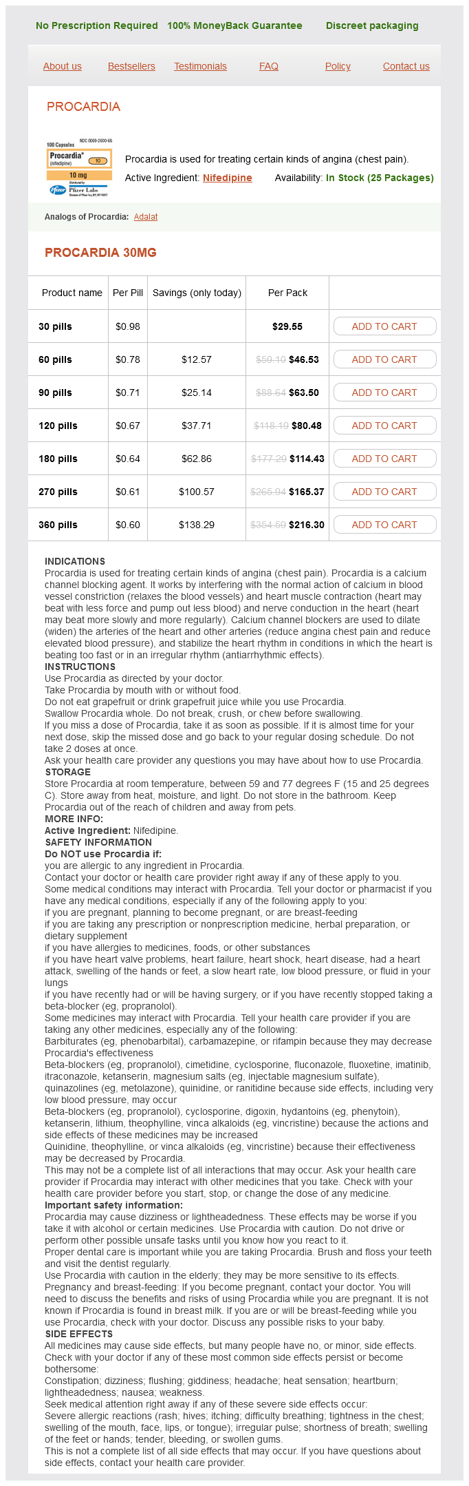
Procardia Dosage and Price
Procardia 30mg
- 30 pills - $29.55
- 60 pills - $46.53
- 90 pills - $63.50
- 120 pills - $80.48
- 180 pills - $114.43
- 270 pills - $165.37
- 360 pills - $216.30
The latter two are associated with a variety of lymphoproliferative or autoimmune disorders and infections arteries of the leg procardia 30 mg mastercard, particularly hepatitis C. Elevated serum levels may be found in inflammatory conditions, renal failure (due to impaired excretion), and in lymphoid and plasma cell malignancies, where it is used as an important marker. Serum levels are not useful to follow chemotherapy responsiveness in myeloma because of the lack of specificity but may predict early relapse following autologous transplantation. There are five distinct isoenzymes, based on different ratios of M and H subunits, with identical function but different distribution; distinguishing between these is seldom useful in hematology practice. Elevated serum levels, which can be measured by an automated enzyme activity-based assay, are seen in tissue damage regardless of etiology, as the enzyme is released from necrotic cells into the circulation. In hematology, marked elevations are usually noted with intravascular or intramarrow hemolysis, hemophagocytic lymphohistiocytosis, and tumor lysis syndrome. Serum Uric Acid Uric acid, the end product of purine nucleotide metabolism in humans, is produced in a variety of cells and cleared by the kidneys. Elevated serum levels (hyperuricemia), as measured by an automated enzymatic assay, are seen with either increased uric acid production (increased cell turnover, as in tumor lysis syndrome) or impaired renal clearance (kidney disease and certain medications). Performance evaluation of the latest fully automated hematology analyzers in a large, commercial laboratory setting: a 4way, side-by-side study. Visual inspection of blood films vs automated analysis of red blood cell distribution width. Quantitative anisocytosis as a discriminant between iron deficiency and thalassemia minor. Relationship of reticulocyte age to polychromasia, shift cells, and shift reticulocytes. Automated reticulocyte counting: state of the art and clinical applications in the evaluation of erythropoiesis. Platelet production and platelet destruction: assessing mechanisms of treatment effect in immune thrombocytopenia. Changes in automated complete blood cell count and differential leukocyte count results induced by storage of blood at room temperature. Bone marrow aspiration before bone marrow core biopsy using the same bone marrow biopsy needle: a good or bad practice A bone marrow report of absent stainable iron is not diagnostic of iron deficiency. Single value of serum transferrin receptor is not diagnostic for the absence of iron stores in anaemic patients with rheumatoid arthritis. Hyperferritinemis is associated with insulin resistance and fatty liver in patients without iron overload. Use of a fixed activated partial thromboplastin time ratio to establish a therapeutic range for unfractionated heparin. Unprovoked recurrent venous thrombosis: prediction by D-Dimer and clinical risk factors. A randomized clinical trial comparing, point-of-care platelet function assays and bleeding time in healthy subjects treated with aspirin or clopidogrel. False-negative results in detection of monoclonal proteins by capillary zone electrophoresis: a prospective study. Highly sensitive, automated immunoassay for immunoglobulin free light chains in serum and urine. Long-term biological variation of, serum protein electrophoresis M-spike, urine M-spike, and monoclonal serum free light chain quantification: implications for monitoring monoclonal gammopathies. Braylan Flow cytometry is a technology used routinely in most hematology laboratories. Its entry into the mainstream of clinical laboratory analysis has been aided by the increasing the availability of monoclonal antibodies that define cell surface and intracellular proteins as markers of cell lineage, differentiation, activation, and other biologic properties. Instrument design advances have yielded benchtop cytometers with fixed optics that, when linked with new developments in fluorochrome chemistry, enable a wide range of clinical applications. In addition, proficiency testing is now available in support of these clinical applications through the College of American Pathologists as mandated by the Clinical Laboratory Improvement Amendment of 1988. The major advantage that flow cytometry provides is its capacity to assess multiple measurements on large numbers of individual cells. Flow cytometric studies have extended the understanding of hematopoietic cell development, differentiation, activation, and apoptosis. In addition, they have provided important information regarding hematologic malignancies, insight into reconstitution after stem cell transplantation, and understanding of cell abnormalities that result in immune or hematologic deficiencies. As such, flow cytometry plays an important role in the diagnosis, characterization, and monitoring of a number of hematologic disorders. The basic design of a flow cytometer involves four major elements: Optics, fluidics, electronics, and a computer equipped with specialized software. At the opposite side of the optical bench, light generated from the cells that have intersected the excitation beam is filtered, reflected by dichroic mirrors set in fixed locations, and finally collected by linked photodetectors to allow quantitation of the emitted light at specific wavelengths. To ensure that all cells analyzed experience consistent exposure to the excitation beam, the fluidic system must maintain the cells in a consistent location as they move sequentially through the beam. To accomplish this, the cell suspension is injected into a flowing stream of sheath fluid that hydrodynamically focuses the inner stream of cells within the outer sheath fluid stream. The various light signals (parameters) are collected by the optical bench, while instrument design determines the number of parameters collected per cell. The two reagent-independent (nonfluorescent) parameters are forward-angle light scatter, as a marker of cell size, and side-angle light scatter, as an index of cellular regularity/ granularity. The combination of these two parameters allows for an approximate discrimination among the three major types of leukocytes, as well as evaluation of red blood cells and platelets in whole blood samples. Fluorochromes are excited by light of a defined wavelength and emit light of lower energy (longer wavelength). There are currently different fluorochromes used in clinical flow cytometry, including fluorescein isothiocyanate, phycoerythrin, peridinin chlorophyll protein, and allophycocyanin and many others that can be excited by newly introduced lasers. Combinations of two fluorochromes linked to each other have been developed; they depend on the transfer of energy from the first fluorochrome to excite the second fluorochrome.