General Information about Nortriptyline
As talked about earlier, nortriptyline is primarily used for the therapy of depression. It has been discovered to be notably effective in people with main depressive disorder, which is characterised by persistent feelings of sadness, hopelessness, and a loss of curiosity in actions that were once gratifying. Nortriptyline may be prescribed for other forms of depression such as bipolar dysfunction, postpartum despair, and seasonal affective disorder.
Before beginning nortriptyline, it could be very important inform the doctor about another medications being taken, together with over-the-counter medicines, as some drugs may interact with nortriptyline and cause antagonistic effects. It can also be recommended to keep away from alcohol consumption while taking nortriptyline, as it might increase the chance of unwanted facet effects.
Nortriptyline works by affecting the balance of neurotransmitters in the brain, particularly norepinephrine and serotonin. These neurotransmitters play an important position in regulating mood and emotions. By increasing their levels, nortriptyline helps to enhance and stabilize a person’s temper, leading to a discount in signs of depression. It may take a number of weeks for the complete effects of nortriptyline to be felt, so it could be very important continue the medication as prescribed by a healthcare skilled.
In conclusion, nortriptyline is a broadly used and efficient treatment for treating melancholy and different psychological health conditions. It has been efficiently used for many years to help people enhance their temper, reduce their symptoms, and lead better lives. While it might cause some side effects, the advantages of nortriptyline far outweigh the potential risks. If you or a liked one are struggling with melancholy, speaking to a healthcare skilled about nortriptyline could presumably be the first step towards improved psychological health and total well-being.
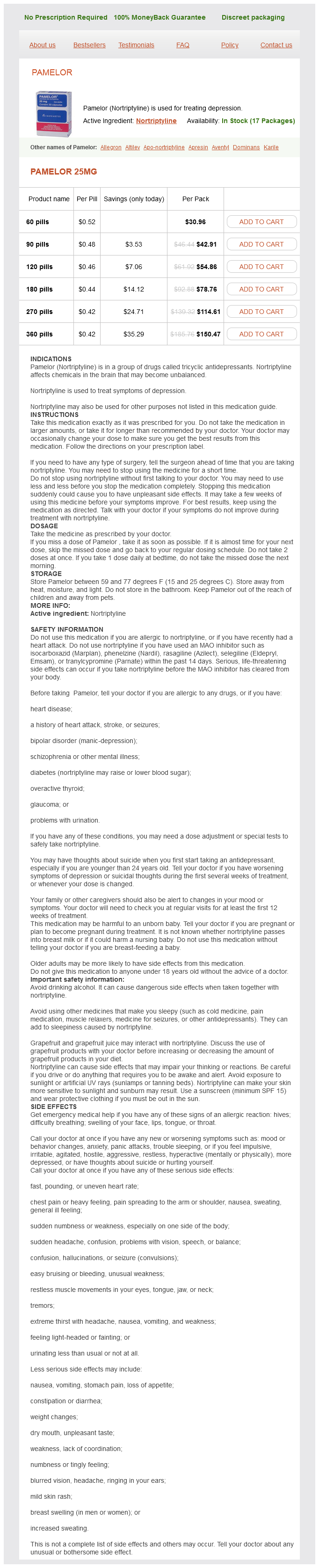
Nortriptyline, generally offered beneath the brand name Pamelor, is a tricyclic antidepressant that has been used for decades to treat varied forms of melancholy. It belongs to a category of medicine known as tricyclic antidepressants, and is known for its effectiveness in bettering mood, lowering anxiousness, and promoting higher sleep. Nortriptyline can be used within the therapy of different psychological health situations corresponding to persistent pain and ADHD. In this article, we'll explore the uses, advantages, and potential side effects of nortriptyline.
Nortriptyline is generally well-tolerated, however as with any treatment, there could also be some unwanted facet effects. The commonest side effects of nortriptyline embody dry mouth, constipation, dizziness, blurred imaginative and prescient, and drowsiness. These side effects normally subside within a couple of weeks of starting the treatment, but if they do not, it may be very important converse to a healthcare skilled. In rarer instances, nortriptyline may trigger more critical side effects similar to low blood stress, critical allergic reactions, and adjustments in heart price. Therefore, it is necessary to often talk with a physician while taking nortriptyline and report any concerning symptoms.
In addition to treating despair, nortriptyline may be prescribed for continual pain situations, corresponding to neuropathic pain and fibromyalgia. Nortriptyline has been found to reduce the depth and frequency of ache, making it a useful possibility for people who don't respond to different ache medicines. It is believed that nortriptyline works by blocking the reuptake of sure chemical substances within the brain that are involved in pain signaling.
The right crus performance anxiety nortriptyline 25 mg order visa, larger and longer than the let crus, arises rom the rst three or our lumbar vertebrae. The thoracic wall and cage have been removed to demonstrate the attachments and convexity o the right dome o the diaphragm. The eshy sternal, costal, and lumbar parts o the diaphragm (outlined with broken lines) attach centrally to the treoil-shaped central tendon, the aponeurotic insertion o the diaphragmatic muscle fbers. The right and let crura and the brous median arcuate ligament, which unites them as it arches over the anterior aspect o the aorta, orm the aortic hiatus. The diaphragm is also attached on each side to the medial and lateral arcuate ligaments. The medial arcuate ligament is a thickening o the ascia covering the psoas major, spanning between the lumbar vertebral bodies and the tip o the transverse process o L1. The lateral arcuate ligament covers the quadratus lumborum muscles, continuing rom the L12 transverse process to the tip o the 12th rib. The superior aspect o the central tendon o the diaphragm is used with the inerior surace o the brous pericardium, the strong, external part o the broserous pericardial sac that encloses the heart. Vessels and Nerves o Diaphragm the arteries o the diaphragm orm a branch-like pattern on both its superior (thoracic) and inerior (abdominal) suraces. The arteries supplying the inerior surace o the diaphragm are the inerior phrenic arteries, which typically are the rst branches o the abdominal aorta; however, they may arise rom the celiac trunk. Some veins rom the posterior curvature o the diaphragm drain into the azygos and hemi-azygos veins (see Chapter 4, Thorax). The veins draining the inerior surace o the diaphragm are the inerior phrenic veins. The lymphatic plexuses on the superior and inerior suraces o the diaphragm communicate reely. The anterior and posterior diaphragmatic lymph nodes are on the superior surace o the diaphragm. Lymph rom these nodes drains into the parasternal, posterior mediastinal, and phrenic lymph nodes. Lymphatic vessels rom the inerior surace o the diaphragm drain into the anterior diaphragmatic, phrenic, and superior lumbar (caval/aortic) lymph nodes. Lymphatic capillaries are dense on the inerior surace o the diaphragm, constituting the primary means or absorption o peritoneal fuid and substances introduced by intraperitoneal (I. Sensory innervation (pain and proprioception) to the diaphragm is also mostly rom the phrenic nerves. Peripheral parts o the diaphragm receive their sensory nerve supply rom the intercostal nerves (lower six or seven) and the subcostal nerves. Also passing through the caval opening are terminal branches o the right phrenic nerve and a ew lymphatic vessels on their way rom the liver to the middle phrenic and mediastinal lymph nodes. Lymphatic vessels are ormed in two plexuses, one on the superior surace o the diaphragm and the other on its inerior surace; the plexuses communicate reely. The phrenic nerves supply all o the motor and most o the sensory innervation to the diaphragm. In most individuals (70%), both margins o the hiatus are ormed by muscular bundles o the right crus. In others (30%), a supercial muscular bundle rom the let crus contributes to the ormation o the right margin o the hiatus. The esophageal hiatus also transmits the anterior and posterior vagal trunks, esophageal branches o the let gastric vessels, and a ew lymphatic vessels. The bers o the right crus o the diaphragm decussate (cross one another) inerior to the hiatus, orming a muscular sphincter or the esophagus that constricts it when the diaphragm contracts. The esophageal hiatus is superior to and to the let o the aortic hiatus is the opening posterior in the diaphragm or the descending aorta. Because the aorta does not pierce the diaphragm, movements o the diaphragm do not aect blood fow through the aorta during respiration. The aorta passes between the crura o the diaphragm posterior to the median arcuate ligament, which is at the level o the inerior border o the T12 vertebra. This triangle transmits lymphatic vessels rom the diaphragmatic surace o the liver and the superior epigastric vessels. The sympathetic trunks pass deep to the medial arcuate ligament, accompanied by the least splanchnic nerves. There are two small apertures in each crus o the diaphragm; one transmits the greater splanchnic nerve and the other the lesser splanchnic nerve. Although this movement is oten described as the "descent o the diaphragm," only the domes o the diaphragm descend. This increases the volume o the thoracic cavity and decreases the intrathoracic pressure, resulting in air being taken into the lungs. In addition, the volume o the abdominal cavity decreases slightly and intra-abdominal pressure increases somewhat. Movements o the diaphragm are also important in circulation because the increased intra-abdominal pressure and decreased intrathoracic pressure help return venous blood to the heart. The diaphragm is at its most superior level when a person is supine (with the upper body lowered, the Trendelenburg position). In this position, the abdominal viscera push the diaphragm superiorly in the thoracic cavity. When a person lies on one side, the hemidiaphragm rises to a more superior level because o the greater push o the viscera on that side. Conversely, the diaphragm assumes an inerior level when a person is sitting or standing. For this reason, people with dyspnea (dicult breathing) preer to sit up, not lie down; nontidal (reserve) lung volume is increased, and the diaphragm is working with gravity rather than opposing it.
A slit-like gap between the medial and the lateral crura o the external oblique aponeurosis anxiety symptoms eye pain purchase generic nortriptyline from india, bridged by intercrural fbers, orms the superfcial inguinal ring. Anterolateral Abdominal Wall 425 the retinaculum spans the subinguinal space, through which pass the fexors o the hip and neurovascular structures serving much o the lower limb. These brous bands are the thickened inerolateral-most portions o the external oblique and aponeurosis and the inerior margin o the transversalis ascia. The inguinal ligament is a dense band constituting the ineriormost part o the external oblique aponeurosis. The most lateral o these bers continue to run along the pecten pubis as the pectineal ligament (o Cooper). Some o the more superior bers an upward, bypassing the pubic tubercle and crossing the linea alba to blend with the lower bers o the contralateral external oblique aponeurosis. The iliopubic tract is the thickened inerior margin o the transversalis ascia, which appears as a brous band running parallel and posterior (deep) to the inguinal ligament. The iliopubic tract, seen in the place o the inguinal ligament when the inguinal region is viewed rom its internal (posterior) aspect. The inguinal ligament and iliopubic tract span and provide central strength to an area o innate weakness in the body wall in the inguinal region called the myopectineal orifce (Fruchaud, 1956). This weak area, occurring in relation to structures traversing the body wall, is the site o direct and indirect inguinal and emoral hernias. It is the beginning o an evagination in the transversalis ascia that orms an opening like the entrance to a cave. Through this opening, the extraperitoneal ductus deerens (vas deerens) and testicular vessels in males (or round ligament o the uterus in emales) and genital branch o the genitoemoral nerve pass to enter the inguinal canal. The transversalis ascia itsel continues into the canal, orming the innermost covering (internal ascia) o the structures traversing the canal. The superfcial (external) inguinal ring is the exit by which the spermatic cord in males, or the round ligament in emales, and ilio-inguinal nerve emerge rom the inguinal canal. The supercial ring is a split that occurs in the diagonal, otherwise parallel bers o the external oblique aponeurosis just superolateral to the pubic tubercle. The parts o the aponeurosis that lie lateral and medial to , and orm the margins o, the supercial ring are crura (L. The lateral crus attaches to the pubic tubercle, and the medial crus attaches to the pubic crest. Fibers o the supercial layer o investing (deep) ascia overlying the external oblique muscle and aponeurosis, running perpendicular to the bers o the aponeurosis, pass rom one crus to the other across the superolateral part o the ring. The inguinal canal is normally collapsed anteroposteriorly against the structures it conveys. Between its two openings (rings), the inguinal canal has two walls (anterior and posterior), as well as a roo and foor. Posterior wall: ormed by the transversalis ascia; its medial part is reinorced by pubic attachments o the internal oblique and transversus abdominis aponeuroses that requently merge to variable extents into a common tendon-the inguinal alx (conjoint tendon)-and the refected inguinal ligament. Roo: ormed laterally by the transversalis ascia, centrally by musculo-aponeurotic arches o the internal oblique and transversus abdominis, and medially by the medial crus o the external oblique aponeurosis. Floor: ormed laterally by the iliopubic tract, centrally by gutter ormed by the inolded inguinal ligament, and medially by the lacunar ligament. The inguinal canal in adults is an oblique passage, approximately 4 cm long, directed ineromedially through the inerior part o the anterolateral abdominal wall. The main occupant o the inguinal canal is the spermatic cord in males and the round ligament o the uterus in emales. These are unctionally and developmentally distinct structures that occur in the same location. The inguinal canal also contains blood and lymphatic vessels, the ilio-inguinal nerve, and the genital branch o the genitoemoral nerve (n. The layers o the abdominal wall and the coverings o the spermatic cord and testis derived rom them are shown. Sagittal section o the anterior abdominal wall and inguinal canal at the plane shown in (A). Most groin hernias in males pass superior to the iliopubic tract (inguinal hernias), whereas most pass inerior to it in emales (emoral hernias). Because o its relative weakness, the myopectineal orice is overlaid with prosthetic mesh placed in the extraperitoneal retro-inguinal space ("space o Bogros") in many hernia repairs. The testes develop in the extraperitoneal connective tissue in the superior lumbar region o the posterior abdominal wall. The male gubernaculum is a brous tract connecting the primordial testis to the anterolateral abdominal wall at the site o the uture deep ring o the inguinal canal. A peritoneal diverticulum, the processus vaginalis, traverses the developing inguinal canal, carrying muscular and ascial layers o the anterolateral abdominal wall beore it as it enters the primordial scrotum. The testis begins to pass through the inguinal canal during the 28th week and takes approximately 3 days to traverse it. As the testis, its duct (ductus deerens), and its vessels and nerves relocate, they are ensheathed by musculoascial extensions o the anterolateral abdominal wall, which account or the presence o their derivatives in the adult scrotum: the internal and external spermatic asciae and cremaster muscle. The stalk o the processus vaginalis normally degenerates; however, its distal saccular part orms the tunica vaginalis, the serous sheath o the testis and epididymis (Moore et al. The ovaries also develop in the superior lumbar region o the posterior abdominal wall and relocate to the lateral wall o the pelvis. The processus vaginalis o the peritoneum traverses the transversalis ascia at the site o the deep inguinal ring, orming the inguinal canal as in the male, and protrudes into the developing labium majus, which is the emale homologue o (part corresponding to) the scrotum. The emale gubernaculum, a brous cord connecting the ovary and primordial uterus to the developing labium majus, is represented postnatally by the ovarian ligament, between the ovary and uterus, and the round ligament o the uterus (L. Because o the attachment o the ovarian ligaments to the uterus, the ovaries do not relocate to the inguinal region; however, the round ligament passes through the inguinal canal and disperses into the subcutaneous tissue o the anterior labium majus. Except or its most inerior part, which becomes a serous sac engulng the testis, the tunica vaginalis, the processus vaginalis obliterates by the 6th month o etal development.
Nortriptyline Dosage and Price
Pamelor 25mg
- 60 pills - $30.96
- 90 pills - $42.91
- 120 pills - $54.86
- 180 pills - $78.76
- 270 pills - $114.61
- 360 pills - $150.47
The valve should be carefully checked for complete freedom of movement of the disk and anxiety symptoms 9 dpo cheapest generic nortriptyline uk, if necessary, the valve is rotated to a point where the greatest clearance from adjacent tissue is achieved. Before completion of the suture line on the atrial septal patch, the left heart is filled with saline, and air is vented through the cardioplegic infusion site in the ascending aorta. The valve is placed entirely within the left atrium between the inferior pulmonary veins and the true annulus. This is usually combined with enlargement of the aortic annulus with the same patch. However, on occasion, we have performed a procedure where the aortic annulus is split between the right and noncoronary leaflets. The aortic valve commissure is reconstructed at the apex of the patch usually with pericardial leaflet extension of the right and noncoronary leaflets to improve aortic valve competence. Although the child was discharged from the hospital, he died 8 months postoperatively. Postoperatively the child remained ventilator dependent and, at catheterization, was found to have what was essentially a ventricular aneurysm because of systolic dilation of the allograft. The right to noncommissure of the aortic valve will be reconstituted at the apex of the triangular prosthetic patch. Supra-annular valve replacement should be applicable in almost all cases in which this procedure might otherwise be contemplated. Results of Surgery Balloon Angioplasty of Congenital Mitral Stenosis One of the first reports of balloon angioplasty for congenital mitral stenosis was published by Spevak et al. In seven of the nine patients effective reduction in mitral gradient was achieved initially. The authors found that surgical resection was preferable in patients with mitral stenosis due to a supravalvar mitral ring. The authors concluded that 5-year survival is relatively poor in patients with severe congenital mitral stenosis, with worse outcomes in infants and patients undergoing valve replacement, but with improvement in the more recent experience. There were three early deaths or transplants with no late deaths over 4 years of follow-up. The report emphasizes the disadvantages of mitral valve replacement in the small infant. Supra-annular mitral valve replacement is often necessary in small babies who cannot avoid replacement. However, subsequent follow-up has suggested that the hemodynamic result with supra-annular mitral valve replacement is suboptimal. It is most commonly found in association with atrioventricular canal (septal) defects where it may be either a preoperative or postoperative problem. The "cleft" of the anterior leaflet is present naturally in the child with a partial atrioventricular canal while, in the child with a complete canal, a cleft is created as part of the repair. Central regurgitation occurs when the valve annulus has dilated to the point where there is poor central apposition of the mural and anterior leaflets. Although the statement is often heard that a child has regurgitation because of "inadequate leaflet tissue," we believe that a more common problem is that repair has been delayed too long allowing ventricular dilation secondary to the volume load of the left to right shunt. Once regurgitation of any cause is present it is likely to worsen over time because the regurgitation per se causes annular dilation resulting in worsening central regurgitation. Furthermore, there are likely to be secondary changes in the valve leaflets such as thickening and rolling of the free edges which will also exacerbate regurgitation. Mitral regurgitation can be a result of structural abnormalities of the valve that are similar to those causing mitral stenosis. Leaflets may be dysplastic and retracted, there may be thick and short chords and the papillary muscles may be abnormal. Other structural abnormalities include isolated cleft of the anterior leaflet, leaflet prolapse secondary to chordal elongation or rupture (usually in the setting of a connective tissue disorder such as Marfan syndrome) or leaflet perforation or other injury by bacterial endocarditis. The symptoms are the usual symptoms of congestive heart failure and are likely to be indistinguishable from the symptoms of mitral stenosis. The degree of mitral regurgitation can be quantitated by assessment of the width of the jet at the level of the leaflets. It is important to do this in two planes as there may be a knife-thin jet coming through the cleft area. As with mitral stenosis, the echocardiographer should analyze the mechanism of the regurgitation. Most commonly this will involve distinguishing regurgitation through the cleft versus central regurgitation. Assessment of the regurgitant mitral valve in the adult with degenerative mitral valve disease is becoming increasingly sophisticated with application of computerized analysis of the various segments of the valve. Because of the variability of congenitally malformed valves this uniform approach to valve description is less useful to the pediatric surgeon than the adult cardiac surgeon. Cardiac catheterization is not necessary or indeed useful in defining the degree or the mechanism of mitral regurgitation. Afterload reduction is particularly helpful in the setting of mitral regurgitation and should be maximized. There are no interventional catheter methods that are useful in the management of mitral regurgitation in the infant or young child. In the adult with acquired mitral regurgitation various innovative catheter methods have been explored, including reshaping the mitral annulus using devices placed in the coronary sinus or annular shrinking with magnets. Indications for Surgery the indications for mitral valve repair for mitral regurgitation should be quite a bit less stringent than those applied for mitral stenosis. This is because it is very likely that the valve can be significantly improved no matter what the cause of the regurgitation. On the other hand, if surgery is delayed there will be secondary changes of the valve which will increase the difficulty of repair and reduce the probability that repair will be successful. It should be highly unlikely that valve replacement is required at a first attempt to improve a regurgitant mitral valve surgically.