General Information about Nasonex nasal spray
Allergies are a standard downside that affects tens of millions of individuals worldwide. Whether it’s the change of seasons or exposure to sure allergens, the signs could be extremely frustrating and disruptive. Nasonex helps present relief from these symptoms, making it a vital treatment for people who undergo from allergies.
Nasonex is a highly effective nasal spray that's used to deal with and forestall nasal signs brought on by allergy symptoms. It is a popular medication prescribed by doctors to help alleviate the discomfort caused by seasonal or year-round allergy symptoms. Nasonex is a kind of corticosteroid, which works by decreasing inflammation within the nasal passages and the release of drugs that set off allergic reactions.
Moreover, Nasonex is a long-lasting treatment that continues to be energetic for as a lot as 24 hours after administration. This signifies that the nasal spray has a sustained impact, making it a more convenient possibility for these with busy schedules. It also reduces the necessity for constant reapplication, making it cheaper in the long run.
Aside from its primary use for relieving allergy signs, Nasonex has additionally been confirmed to be efficient in preventing future symptoms. It works by reducing irritation within the nasal passages and blocking the release of substances that trigger allergic reactions. This is especially helpful for individuals who experience allergies year-round and wish to proactively manage their symptoms.
Another important benefit of using Nasonex is its safety and low risk of unwanted effects. Unlike many other allergy medications, Nasonex is applied directly to the affected area, minimizing potential unwanted effects such as drowsiness and dry mouth that can occur with oral medication. When used as directed, Nasonex is considered secure and well-tolerated. However, like several medicine, there's still a small probability of unwanted side effects, and it is important to seek the guidance of a doctor before use, particularly if there are pre-existing medical circumstances.
In conclusion, Nasonex is a wonderful option for those in search of aid from allergy signs. Its ease of use, fast motion, and long-lasting impact make it a preferred selection amongst each docs and sufferers. It is necessary to notice that Nasonex is a prescription medication and will only be used under the steering of a healthcare skilled. If you would possibly be struggling with nasal symptoms brought on by allergic reactions, converse to your doctor right now to see if Nasonex could also be an appropriate remedy possibility for you.
One of essentially the most important advantages of Nasonex is its ease of use. It comes in the form of a nasal spray, making it painless and convenient to manage. There isn't any need for injections or drugs, making it an excellent possibility for these who dislike taking medication orally or are afraid of needles. The nasal spray can be simply carried around and used each time needed, making it a convenient resolution for folks on the go.
Nasonex can be recognized for its rapid onset of motion. Unlike oral treatment that may take a while to work, the nasal spray directly targets the nasal passages, providing almost immediate aid. The lively ingredient in Nasonex, mometasone furoate, has been confirmed to work quickly in decreasing nasal inflammation and relieving signs like congestion, sneezing, and a runny nostril.
The association of knee injury and obesity with unilateral and bilateral osteoarthritis of the knee allergy shots vancouver bc buy generic nasonex nasal spray. Comparison of radiological osteoarthritis in a Dutch population with that in 10 other populations. A meta-analysis of sex differences prevalence, incidence and severity of osteoarthritis. Incidence and progression of osteoarthritis in women with unilateral knee disease in the general population: the effect of obesity. Life course body mass index and risk of knee osteoarthritis at the age of 53 years: evidence from the 1946 British birth cohort study. The longitudinal relationship between changes in body weight and changes in medial tibial cartilage, and pain among community-based adults with and without meniscal tears. Prevalence of hip symptoms and radiographic and symptomatic hip osteoarthritis in African Americans and Caucasians: the Johnston County Osteoarthritis Project. Differences in radiographic features of knee osteoarthritis in African-Americans and Caucasians: the Johnston county osteoarthritis project. Occurrence of radiographic osteoarthritis of the knee and hip among African Americans and whites: a population-based prospective cohort study. The relationship between osteoarthritis and osteoporosis in the general population: the Chingford Study. Association of radiographically evident osteoarthritis with higher bone mineral density and increased bone loss with age. Axial and hip bone mineral density and radiographic changes of osteoarthritis of the knee: data from the Baltimore Longitudinal Study of Aging. Bone mineral density and risk of incident and progressive radiographic knee osteoarthritis in women: the Framingham Study. Bone mineral density and vertebral fracture history are associated with incident and progressive radiographic knee osteoarthritis in elderly men and women: the Rotterdam Study. The relationship of bone density and fracture to incident and progressive radiographic osteoarthritis of the knee: the Chingford Study. Cross-sectional and longitudinal associations between systemic, subchondral bone mineral density and knee cartilage thickness in older adults with or without radiographic osteoarthritis. Occupational physical demands, knee bending, and knee osteoarthritis: results from the Framingham Study. Habitual physical activity is not associated with knee osteoarthritis: the Framingham Study. Mechanical and constitutional risk factors for symptomatic knee osteoarthritis: differences between medial tibiofemoral and patellofemoral disease. Factors associated with radiographic osteoarthritis: results from the population study 70-year-old people in Goteborg. Effect of recreational physical activities on the development of knee osteoarthritis in older adults of different weights: the Framingham Study. Meeting physical activity guidelines and the risk of incident knee osteoarthritis: a population-based prospective cohort study. No Association between Daily Walking and Knee Structural Changes in People at Risk of or with Mild Knee Osteoarthritis. Level of physical activity and the risk of radiographic and symptomatic knee osteoarthritis in the elderly: the Framingham study. High plasma levels of vitamin C and E are associated with incident radiographic knee osteoarthritis. The association between vitamin K status and knee osteoarthritis features in older adults: the Health, Aging and Body Composition Study. Relation of dietary intake and serum levels of vitamin D to progression of osteoarthritis of the knee among participants in the Framingham Study. Milk consumption and progression of medial tibiofemoral knee osteoarthritis: data from the Osteoarthritis Initiative. Moderate vitamin D deficiency is associated with changes in knee and hip pain in older adults: a 5-year longitudinal study. Effect of vitamin D supplementation on progression of knee pain and cartilage volume loss in patients with symptomatic osteoarthritis: a randomized controlled trial. Effect of Vitamin D, Supplementation on Tibial Cartilage Volume and Knee Pain Among Patients With Symptomatic Knee Osteoarthritis: A Randomized Clinical Trial. Cigarette smoking and risk of osteoarthritis in women in the general population: the Chingford study. The relationship between smoking and knee osteoarthritis in the Osteoarthritis Initiative. History, of knee injuries and knee osteoarthritis: a meta-analysis of observational studies. Association of knee injuries with accelerated knee osteoarthritis progression: data from the Osteoarthritis Initiative. The natural history of bone marrow lesions in community-based adults with no clinical knee osteoarthritis. Meniscal tear in knees without surgery and the development of radiographic osteoarthritis among middle-aged and elderly persons: the Multicenter Osteoarthritis Study. Significance of preradiographic magnetic resonance imaging lesions in persons at increased risk of knee osteoarthritis. Multitissue Involvement Leading to Radiographic Osteoarthritis: Magnetic Resonance Imaging-Based Trajectory Analysis Over Four Years in the Osteoarthritis Initiative. Patterns of Coexisting Lesions Detected on Magnetic Resonance Imaging and Relationship to Incident Knee Osteoarthritis: the Multicenter Osteoarthritis Study. Relationship of, meniscal damage, meniscal extrusion, malalignment, and joint laxity to subsequent cartilage loss in osteoarthritic knees.
Fascicles of the thoracic component of longissimus thoracis (longissimus thoracis pars thoracis) arise from all thoracic transverse processes and most ribs allergy shots eczema cheap 18 gm nasonex nasal spray with visa, and attach to either lumbar spinous processes, the sacrum, or the ilium. For example, instead of distinct tendinous attachments to bone, many spinal muscles have very little tendon at their ends, but have a complex arrangement of internal tendons and aponeuroses. Some spinal muscles have short fascicles and high pennation, whereas others have long, parallel fascicles. All of these factors afect the force- and moment-generating capacity of muscles (as described in Chapter 3), which ultimately inluences control of spinal movement and injury mechanisms. Spinal muscles can be divided into intrinsic muscles, which connect vertebrae with each other or with the skull, and extrinsic muscles, which attach vertebrae to the limbs, shoulder girdle, ribcage, or pelvis. Embryologically, intrinsic muscles originate from the epimere and extrinsic muscles originate from the hypomere. Intrinsic muscles receive innervation from the dorsal rami of spinal nerves, whereas extrinsic muscles are innervated by the ventral rami of spinal nerves and generally have functions related more to the proximal portion of limbs or respiration. Intrinsic Spinal Muscles in the Lumbar, Thoracic, or Cervical Spine Intrinsic muscles of the spine are dominated by the erector spinae, a group of interdigitated muscles that span the entire length of the spine, from the sacrum and iliac crest to the skull. Another important group of muscles, the multiidus, are shorter and deeper, and are described in more detail later. In the thoracolumbar region, the erector spinae and multiidus muscles comprise the bulk of the spinal musculature. Architectural analysis and intraoperative measurements demonstrate the unique design of the multiidus for lumbar spine stability. In contrast to the more medially located longissimus thoracis, the caudal tendons are less prominent, giving the iliocostalis lumborum a much more leshy appearance. Caudad to rib 10, the iliocostalis lumborum and longissimus thoracis lie side by side, forming the erector spinae aponeurosis. Rostral to rib 9 or 10, the iliocostalis thoracis separates the iliocostalis lumborum and longissimus thoracis. In the upper thoracic and cervical region, the iliocostalis cervicis connects the ribs to the transverse processes of cervical vertebrae. MacIntosh and Bogduk measured muscle and tendon lengths in the thoracic portions of the longissimus thoracis and iliocostalis lumborum (Table 4. Underneath the trapezius lie the splenius capitis, splenius cervicis, levator scapulae, and rhomboids. Left side shows semispinalis cervicis and the suboccipital muscles, which lie under semispinalis capitis. Because thoracic fascicles of the longissimus thoracis and iliocostalis lumborum lie over the lumbar fascicles, electrodes placed at lumbar vertebral levels may not represent activity of fascicles directly attached to lumbar vertebrae. Multiidus muscles arising from lower lumbar vertebrae consist of fewer fascicles because the number of vertebrae caudad to the origin decreases. Its iber type distribution of approximately 60% type 1 ibers supports this postural role. In fact, similar iber type distributions between multiidus and erector spinae muscles suggest similar functions for these two muscle groups. Second, direct mechanical testing of the multiidus muscle cells and extracellular connective tissue revealed that, while the multiidus ibers have the same mechanical properties as limb muscles, the iber bundles, which include extracellular connective tissue, are about twice as stif. In other words, as the spine lexes, multiidus force increases, suiting it to restore spine angles toward neutral or more extended positions. Deep to the multiidus are smaller muscles that span one or two vertebral segments. Rotators are prominent in the thoracic region; although some authors claim that they exist in the lumbar region,11,12 MacIntosh and Bogduk did not ind any muscles deep to the lumbar multiidus. Intrinsic Spinal Muscles Speciic to the Cervical Spine Because of diferent functional demands in the cervical spine. Kamibayashi and Richmond quantiied neck muscle anatomy and architecture of the neck muscles (Table 4. Contiguous, slightly deeper, and sometimes inseparable is the splenius cervicis, which originates on thoracic spinous processes and inserts on cervical transverse processes. Although both the splenius capitis and splenius cervicis function in extension, lateral bending, and axial rotation, the splenius capitis is oriented more obliquely than the splenius cervicis, providing more axial rotation capacity for movements of the skull relative to the vertebrae. The medial portion is characterized by tendinous inscriptions and internal aponeuroses interrupting the fascicles. Because it lies close to the vertebral bodies, it has only a small lexion moment arm; the superomedial orientation could provide ipsilateral rotation. Note the three parts of longus colli: superior oblique, vertical, and inferior oblique. All four of these muscles can contribute to extension of the head with respect to the neck. In addition, the rectus capitis posterior major and the obliquus capitis inferior are oriented to produce ipsilateral rotation, and the lateral location of the obliquus capitis superior implies a lateral bending function. Fascicles of the psoas generally have the same length, regardless of their level of origin. Electromyographic evidence shows that the quadratus lumborum has a dominant role in spine stabilization. Kamibayashi and Richmond4 divided this muscle into three subvolumes: sternomastoid, cleidomastoid, and cleido-occipital. Arrows indicate diferences in pulling direction of deep (D) and supericial (S) subvolumes. It can be divided into three segments: the rostral segment (also called clavotrapezius or trapezius pars descendens) runs from the lateral part of the clavicle to the occiput or ligamentum nuchae; the middle part (acromiotrapezius or pars transversa) runs nearly perpendicular to the midline at the lower cervical and upper thoracic levels from the lateral part of the scapular spine; and the caudal part (spinotrapezius or pars ascendens) attaches to spinous processes of T4 to T12 from the scapula.
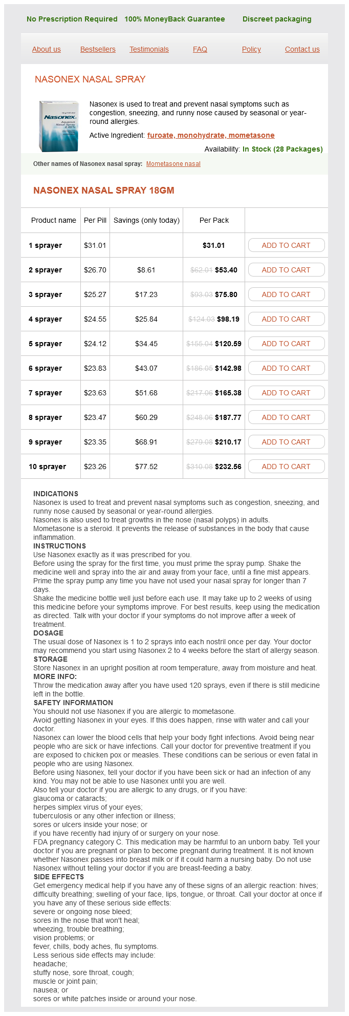
Nasonex nasal spray Dosage and Price
Nasonex nasal spray 18gm
- 1 sprayer - $31.01
- 2 sprayer - $53.40
- 3 sprayer - $75.80
- 4 sprayer - $98.19
- 5 sprayer - $120.59
- 6 sprayer - $142.98
- 7 sprayer - $165.38
- 8 sprayer - $187.77
- 9 sprayer - $210.17
- 10 sprayer - $232.56
With such typical features allergy treatment center st louis order genuine nasonex nasal spray on-line, a confident clinical diagnosis can be made in adults aged >40 yr. Clinical hallmarks: Heberden and Bouchard nodes and/or bony enlargement with or without deformity. Function should be carefully assessed and monitored using validated outcome measures. A posteroanterior radiograph of both hands on a single film or field of view is adequate for diagnosis. It is a common complex joint disorder showing focal cartilage loss, new bone formation, and involvement of all joint tissues. However, the ability to discriminate subsets and the relevance for routine practice are unclear. Additional features that may be present include deformity (fixed flexion and/or varus-less commonly valgus), instability, periarticular or joint-line tenderness, and pain on patellofemoral compression. Other important considerations are referred pain, ligamentous and meniscal lesions, and bursitis. Classical features are focal joint space narrowing, osteophyte, subchondral bone sclerosis, and subchondral cysts. If a palpable effusion is present, synovial fluid should be aspirated and analyzed to exclude inflammatory disease and to identify urate and calcium pyrophosphate crystals. Plain radiography and magnetic resonance imaging diagnostics in osteoarthritis: validated staging and scoring. Prevalence, incidence, and progression of hand osteoarthritis in the general population: the Framingham Osteoarthritis Study. Because many factors such as country of residence, health care system factors, demographic factors, comorbidities, willingness to undergo surgery, and psychological factors influence the receipt of a joint replacement, perhaps a more appropriate metric in this regard would be progression leading to the need for total joint replacement; how this would be defined for research and policy purposes is currently the subject of considerable attention in the epidemiologic research community. However, recent studies using novel methods to control for confounding have suggested a closer relationship between radiographic findings and symptoms. In the Study of Osteoporotic Fractures, of approximately 6000 women followed over 8 years, 12. Although a variety of confounding factors contribute to whether or not an individual gets a joint replacement. The effect of osteoarthritis definition on prevalence and incidence estimates: a systematic review. The American College of Rheumatology criteria for the classification and reporting of osteoarthritis of the hip. Impact of different descriptions of the Kellgren and Lawrence classification criteria on the diagnosis of knee osteoarthritis. The natural history of radiographic knee osteoarthritis: A fourteen-year population-based cohort study. Defining incident radiographic hip osteoarthritis for epidemiologic studies in women. Prevalence of radiographic and symptomatic hip osteoarthritis in an urban United States community: the Framingham osteoarthritis study. Extreme variations in racial rates of total hip arthroplasty for primary coxarthrosis: a population-based study in San Francisco. Very low prevalence of hip osteoarthritis among Chinese elderly in Beijing, China, compared with whites in the United States: the Beijing osteoarthritis study. Prevalence and Risk Factors of Spine, Shoulder, Hand, Hip, and Knee Osteoarthritis in Community-dwelling Koreans Older Than Age 65 Years. Prevalence of knee, osteoarthritis in the United States: arthritis data from the Third National Health and Nutrition Examination Survey 1991-94. Increasing prevalence of knee pain and symptomatic knee osteoarthritis: survey and cohort data. Prevalence of knee symptoms and radiographic and symptomatic knee osteoarthritis in African Americans and Caucasians: the Johnston County Osteoarthritis Project. Prevalence of knee osteoarthritis, risk factors, and quality of life: the Fifth Korean National Health And Nutrition Examination Survey. Comparison of the prevalence of knee osteoarthritis between the elderly Chinese population in Beijing and whites in the United States: the Beijing Osteoarthritis Study. Case definitions of knee osteoarthritis in 4,151 unselected subjects: relevance for epidemiological studies: the Copenhagen Osteoarthritis Study. Joint space, width of the tibiofemoral and of the patellofemoral joint in chronic knee pain with or without radiographic osteoarthritis: a 2-year follow-up. Prevalence and pattern of radiographic hand osteoarthritis and association with pain and disability (the Rotterdam study). Prevalence, incidence and progression of hand osteoarthritis in the general population: the Framingham Osteoarthritis Study. Radiographically defined osteoarthritis of the hand and knee in young and middle-aged African American and Caucasian women. Differences in multijoint radiographic osteoarthritis phenotypes among African Americans and Caucasians: the Johnston County Osteoarthritis project. Lower prevalence of hand osteoarthritis among Chinese subjects in Beijing compared with white subjects in the United States: the Beijing Osteoarthritis Study. Comparison of the prevalence of radiographic osteoarthritis of the knee and hand between Japan and the United States. Lumbar spine radiographic features and demographic, clinical, and radiographic knee, hip, and hand osteoarthritis. Radiographic evaluation of foot osteoarthritis: sensitivity of radiographic variables and relationship to symptoms. Methodologic challenges in studying risk factors for progression of knee osteoarthritis. Association of incident, symptomatic hip osteoarthritis with differences in hip shape by active shape modeling: the Johnston County Osteoarthritis Project.