General Information about Himcolin
Erectile dysfunction is a standard condition that affects hundreds of thousands of males worldwide. It is characterised by the lack to achieve or keep an erection throughout sexual exercise. This can have a significant impression on one's confidence, self-esteem, and general high quality of life. While there are various treatment choices out there, many men are turning to Himcolin gel as a protected and natural answer for his or her sexual woes.
Apart from its main perform of improving erectile perform, Himcolin gel additionally has different benefits for males's sexual health. It helps in sustaining overall penile well being, prevents premature ejaculation, and may even improve the scale of the penis with common use. These added advantages make it an all-in-one answer for males battling sexual problems.
Another necessary ingredient in Himcolin gel is Hygrophila, which is understood for its aphrodisiac properties. It helps in enhancing libido and enhancing sexual efficiency. This herb has been utilized in Ayurvedic medication to treat male sexual dysfunction, and its inclusion in this gel is a testomony to its effectiveness.
The key elements in Himcolin gel work collectively to stimulate blood move to the genitals, leading to harder and long-lasting erections. One of the principle components within the gel is the herb, Celastrus paniculatus, which has been utilized in traditional Indian medication for hundreds of years to deal with male sexual problems. It has been found to enhance the manufacturing of nitric oxide, a compound that helps chill out blood vessels and will increase blood circulate to the penis.
Himcolin gel is a novel and effective product that has been particularly designed to assist males achieve long-lasting erections. This remarkable gel is created from natural and herbal ingredients, which makes it protected and free from unwanted effects. Its powerful formulation and ability to enhance sexual efficiency has made it a well-liked selection amongst men seeking a solution for erectile dysfunction.
Himcolin gel also incorporates different potent herbs such as Musk, Nutmeg, and Almond, which work together to enhance blood circulation and improve sensitivity within the genitals. These herbs not solely improve sexual function but additionally have a calming impact on the mind and physique, which can help alleviate performance-related anxiousness and stress.
Many males have reported a big enchancment in their erectile perform after utilizing Himcolin gel. It is easy to apply and begins to work inside minutes, making it a convenient and efficient answer for men who wish to boost their intercourse life. The no-mess formulation of the gel also makes it discreet, and it could be simply carried in a pocket or bag for spontaneous moments of intimacy.
In conclusion, Himcolin gel is a secure and efficient resolution for men who need to obtain long-lasting erections. Its pure and herbal elements make it a viable possibility for people who choose to avoid chemical-based medications. Regular use of this gel can't solely improve sexual perform, but also increase confidence and result in a healthier and happier intercourse life. However, like some other treatment, it is suggested to seek the guidance of a doctor earlier than utilizing Himcolin gel, particularly when you have any underlying medical conditions.
Although lipodystrophia centrifugalis abdominalis infantilis affects primarily Asian children erectile dysfunction drugs forum buy himcolin 30 gm free shipping, it may occasionally occur in Caucasians and in adults. Clinically, a well-demarcated area of lipoatrophy with a periphery of erythema and scale is seen on the abdomen or elsewhere on the trunk, often with associated regional lymphadenopathy. The underlying blood vessels become visible and ulceration may occur within the depressed areas. The lesions may progress slowly over several years, then often cease enlarging by 13 years of age52. Progressive hemifacial atrophy (ParryRomberg syndrome) is considered to be a form of severe morphea (see Ch. As with insulin lipoatrophy, anti-insulin antibodies are associated with lipohypertrophy in children and adolescents with type 1 diabetes. The clinical consequence is that injection of insulin into a site of lipohypertrophy, while painless, may lead to erratic absorption of the insulin, with the potential for poor glycemic control. Of note, insulin-induced nodular amyloidosis may have a clinical appearance similar to lipohypertrophy. Injection of pegvisomant, a growth hormone receptor antagonist used to treat acromegaly, is another cause of lipohypertrophy (Table 101. Non-progressive late-onset linear hemifacial lipoatrophy occurs on the malar cheek, primarily in elderly individuals. Inflammatory reactions due to material injected for cosmetic purposes or from self-inflicted injections of foreign material can lead to lipoatrophy which may be associated with scarring and dyschromia. Less often, biopsy specimens appear to have no overt abnormalities, except for an increased amount of collagen, which has replaced the subcutaneous fat. In acquired generalized lipodystrophy and areas of lipoatrophy in some forms of partial lipodystrophy, two histologic patterns have been recognized: (1) non-inflammatory with involutional changes of the fat; and (2) inflammatory in the form of a lobular panniculitis with lymphocytes, lipophages, and plasma cells55. Whether these two patterns are stage-related remains to be clarified, as even in patients without panniculitis clinically, biopsies of early lesions may show inflammation, but it is usually mild. In the first type, the lobules may be composed of faintly acidophilic, small fat cells that retract from one another as well as the surrounding connective tissue, leading to an appearance similar to embryonic fat; the lobules often have an eosinophilic appearance. These histologic findings are more prominent at the periphery of the clinically depressed area, with tiny acidophilic fat cells present more centrally. The second type, with indistinguishable clinical features, shows small atrophic adipocytes with a normal fat cell membrane surrounded by prominent vasculature. At scanning magnification, the lobules are collapsed (with an orientation parallel to the skin surface) and surrounded by numerous capillaries. Acid mucopolysaccharide deposition within the fat lobules and fibrosis of the septae may be seen, as well as occasionally lipomembranous changes. Although birefringent, non-crystalline foreign material may be observed, foreign body giant cells and lipophages are typically absent in both subtypes of involutional lipoatrophy. Ultrastructural studies have shown that these are lysosomally active macrophages adjacent to lipocytes, and they may contain degenerated lipid38. In localized lipodystrophy, a loss of fatty tissue is seen, along with a variable degree of fibrosis. There is usually no inflammatory infiltrate, although panniculitis has been reported in a few cases. Deposits of immunoreactants can be seen within blood vessel walls or the basement membrane, particularly in the inflammatory type. Histologically, well-developed areas of lipodystrophia centrifugalis abdominalis infantilis show a diminution of subcutaneous fat with scant or absent inflammation. The erythematous rim tends to correspond to a moderate or marked lymphohistiocytic infiltrate in the subcutaneous fat; a deep dermal and peri-eccrine lymphocytic infiltrate is an uncommon finding. Other localized variants such as lipoatrophia semicircularis (semicircular lipoatrophy) and drug-induced lipoatrophy usually show a loss of fatty tissue and replacement by collagen, with no features of panniculitis. However, at sites of glatiramer acetate injections, panniculitis that resembles lupus profundus histologically can be seen. In an infant with generalized lipodystrophy, the following conditions should also be considered: Leprechaunism (Donohue syndrome) is included in some lipodystrophy classifications, as the patients present with generalized lipodystrophy, severe insulin resistance, acanthosis nigricans, and hirsutism. Progeria-type syndromes are characterized by limb lipoatrophy, cardiovascular disease and diabetes, but they are accompanied by muscle wasting, sclerodermatous changes, cataracts, and other signs of premature aging. In the case of partial lipodystrophy, there was a report of diffuse, symmetrical lipoatrophy of the lower extremities following extensive inflammation due to lobular panniculitis56. Localized lipodystrophies should be differentiated from initial phases of progressive lipodystrophy, morphea, lupus panniculitis, and atrophoderma of Pasini and Pierini. Although there is overlap amongst the various types of localized lipodystrophy, the distribution and morphology combined with a history of injections, the presence clinically or histologically of panniculitis, and associated autoimmune disorders may aid in the diagnosis. Poland syndrome is a rare congenital disorder consisting of unilateral partial or total absence of a breast and/or pectoralis major muscle plus ipsilateral symbrachydactyly, which may simulate lipoatrophy. In trauma-induced cases, in particular lipoatrophia semicircularis, the depressions may normalize over a period of weeks. Switching to purified human insulin may improve insulin lipoatrophy, but it may take 13 years54. Soft tissue augmentation using a variety of permanent and non-permanent fillers may be helpful (see Ch. Solid synthetic volumetric midfacial implants, constructed using computer-aided design and manufacturing technology57, may offer a more durable correction of the defects. A thoughtful assessment of possible long-term complications of permanent fillers is always mandatory prior to treatment. While medical therapy is directed predominantly at the metabolic derangements in order to reduce the associated morbidity and mortality, it may also offer some benefit for the lipodystrophic features. However, there have been concerns regarding the adverse side effects of this group of drugs, including hepatotoxicity, weight gain, congestive heart failure, and bone fractures in women.
Resolution of the illness usually occurs after 2 to 3 weeks erectile dysfunction young cure cheap 30 gm himcolin free shipping, although symptoms of fatigue and malaise may last longer. Heterophile antibodies are capable of agglutinating sheep, horse, or bovine erythrocytes, and they typically become detectable 1 week to 1 month after the onset of symptoms and persist for 3 to 18 months. Administration of ampicillin or amoxicillin, and less commonly penicillin or cephalosporins, to patients with infectious mononucleosis, frequently (up to 90% with the former agents) leads to the development of a "hypersensitivity" skin reaction. In the absence of a pre-existing allergy, such reactions do not generally preclude the use of this class of antibiotics after the infectious mononucleosis resolves. Significant enlargement of oropharyngeal lymphoid tissue can lead to airway compromise. A combination of history, physical, examination, and laboratory studies can differentiate among these diseases. Treatment In most cases of infectious mononucleosis, the disease is self-limited and treatment is supportive. Due to their multiple side effects, corticosteroids are reserved for only complicated cases of infectious mononucleosis associated with hemolytic anemia, severe thrombocytopenia, liver failure, or lymphadenopathy resulting in airway compromise62. To decrease this risk, leukocytedepleted blood can be used in immunosuppressed patients. It is a cytotoxic virus that causes cell enlargement (cytomegaly) and nuclear condensation (inclusion). In immunocompromised individuals, however, reactivation of latent virus can lead to recurrent infection characterized by persistent viral replication, viremia, and dissemination to distant organs. Cell injury and tissue destruction contribute to organ damage by directly altering cellular function and producing an inflammatory response. Infection in healthy children and adults often involves the lymphoid tissues, while fetal and neonatal infection commonly affects the salivary glands and neurons. Infection in immunocompromised individuals frequently involves the retina and lungs as well as the liver and gastrointestinal tract. This syndrome has also been described in both immunocompetent and immunocompromised individuals after a blood transfusion. Infected patients develop non-exudative pharyngitis in addition to fever, malaise, myalgias, lymphadenopathy, and hepatosplenomegaly. An exanthem, which can be morbilliform, urticarial, petechial or purpuric, develops in a small percentage of patients. As with infectious mononucleosis, the administration of ampicillin or related drugs during this symptomatic period leads to a cutaneous eruption in 80100% of individuals. Approximately 510% of infected neonates have symptoms at birth, presenting with jaundice, hepatosplenomegaly, intrauterine growth retardation, thrombocytopenia, chorioretinitis, seizures, and/or intracranial calcifications. Cutaneous manifestations include purpuric papules and nodules of dermal History In 1881, Ribbert first observed "protozoan-like cells" in the organs of a stillborn baby with presumed congenital syphilis. Before the discovery of a viral etiology, the disease was initially called "cytomegalic inclusion disease" based on the characteristic enlarged cells (cytomegalia) and intranuclear inclusions. These cytomegalic cells are most likely to be evident in ulcers, and vascular dilatation is another common finding. Treatment and Prevention Management is targeted at prevention plus prophylactic antiviral treatment in susceptible immunocompromised individuals as well as antiviral treatment in those who are symptomatic. When patients fail to respond to these medications or develop significant side effects, foscarnet and cidofovir are additional options69 (Table 80. Despite the high seroprevalence, only 30% of children develop clinical manifestations of exanthem subitum. It has been postulated that drug-related immunosuppression or induction of viral replication leads to viral reactivation, which stimulates a cellular immune response directed against herpesviruses and likely cross-reacting with the drug. Typically, an otherwise well-appearing infant presents with a high fever (102105°F; 38. The cutaneous eruption usually lasts for 2448 hours and may develop before the temperature returns to normal or up to 2 days afterwards. The lesions are discrete, circular or elliptical, "rose-red" macules and papules that are 25 mm in diameter and occasionally surrounded by a white halo. Eyelid edema, mild upper respiratory symptoms, injection of the tympanic membranes, cervical or occipital lymphadenopathy, and a bulging anterior fontanelle may also be evident. The most common complication of exanthem subitum is febrile seizures, which occur in 1015% of affected infants and children. History Zahorsky distinguished exanthem subitum from other exanthems in 1910 and is credited for its first description. Infection is most common in young children, occurring between the ages of 6 months and 3 years in 95% of cases, with a peak incidence between 6 and 12 months of age. The presence of IgM antibodies indicates primary infection, but this assay is not reliable for the detection of reactivation. Histopathologically, exanthem subitum shows nonspecific changes such as spongiosis of the epidermis with spongiform vesicles and exocytosis of lymphocytes. A sparse inflammatory infiltrate is found in the superficial dermis, and papillary dermal edema is often evident. Other considerations may include scarlet fever, Rocky Mountain spotted fever, and Kawasaki disease (see Ch. Evidence-based recommendations regarding specific antiviral agents and dosages are not currently available to guide treatment in immunocompromised hosts. Expansion in the number of immunocompromised patients has led to significant increases in the diversity and severity of clinical presentations of human herpesvirus infections. Thus, it is imperative to be aware of the various manifestations of these viral infections and to perform appropriate diagnostic tests. The clinician must also be aware of the indications, contraindications, dosages, and limitations of antiviral agents.
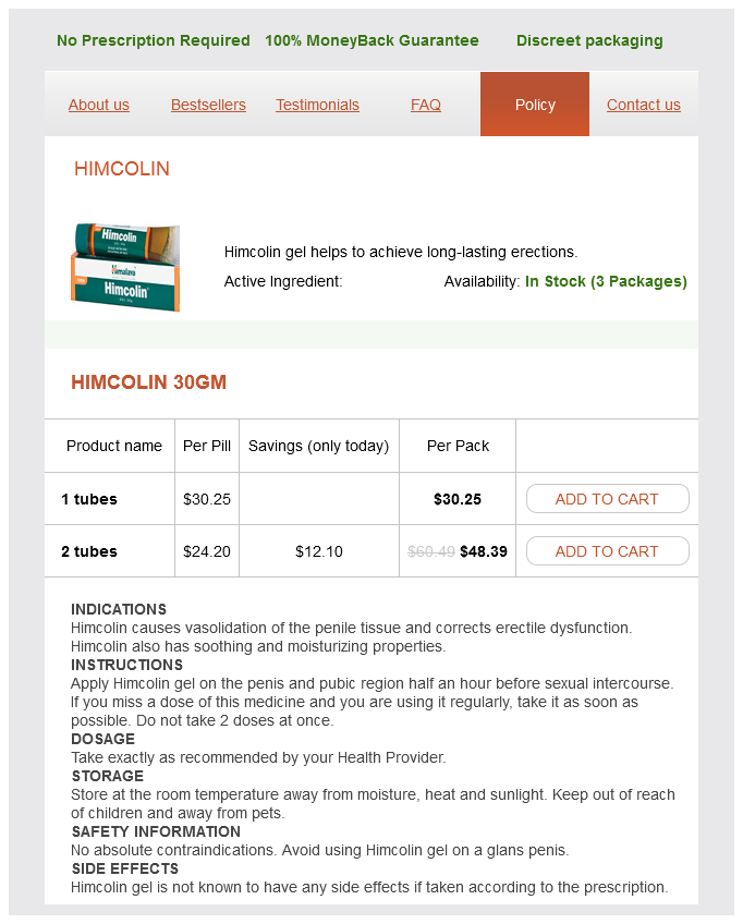
Himcolin Dosage and Price
Himcolin 30gm
- 1 tubes - $30.25
- 2 tubes - $48.39
Dermoid Cyst Cutaneous dermoid cysts typically present in an infant along an embryonic fusion plane as a discrete erectile dysfunction treatment injection cheap himcolin online visa, subcutaneous nodule (see Ch. Although most ear pits are incidental findings, in a newborn, a physical examination to exclude one of the associated syndromes and an evaluation for hearing loss is indicated. Pathology Histologically, dermoid cysts are lined by stratified squamous epithelium that includes a granular layer. They contain other normal cutaneous structures such as hair, sebaceous lobules, eccrine glands, apocrine glands, and/or smooth muscle. Pilonidal cysts typically present as an inflamed, painful, cystic swelling in the upper gluteal cleft or sacrococcygeal area, but they have been described in other locations. Numerous,tiny, translucentorbluish papulesonthelower eyelid(A)orthecheek (B) and in males who are hirsute21. Some authors have argued that they are congenital, essentially representing a dermoid cyst, whereas most authors now believe that the vast majority of lesions are acquired, representing a foreign body response to entrapped hair. A pilonidal cyst can be seen as part of the "follicular inclusion tetrad" which consists of acne conglobata, hidradenitis suppurativa, dissecting cellulitis (perifolliculitis capitis abscedens et suffodiens), and pilonidal cyst (see Ch. Persistent exogenous hairs in the interdigital space of barbers or dog groomers may incite an encompassing epidermal proliferation, giving rise to a pilonidal cyst. Pathology Histologic features are those of an epidermal-lined cyst or sinus tract. Cyst cavities contain hair and keratin debris and are surrounded by granulation tissue and mixed inflammation. Hidrocystomas are traditionally divided into apocrine and eccrine hidrocystomas by histologic features, and as solitary (Smith type) or multiple(Robinson type). Hidrocystomas may be associated with specific syndromes of ectodermal dysplasia, including SchöpfSchulzPassarge syndrome. Eccrine hidrocystomas can enlarge with heat exposure or during the summer and regress with cooler temperatures. In general, eccrine hidrocystomas are thought to develop from cystic dilation of eccrine ducts due to retention of eccrine secretions, while apocrine hidrocystomas are thought to represent adenomas of apocrine sweat gland coils22. Apocrine hidrocystomas are sometimes referred to as cystadenomas, although it has been recommended that this term be reserved for lesions with true papillomatous projections histologically23. Hidrocystomas that appear by Treatment Hidrocystomas may be removed by simple excision, including via Gradle scissors, or electrodesiccation. Multiple eccrine hidrocystomas may also be treated with daily application of topical 1% atropine in aqueous solution, although lesions reappear within days of discontinuing therapy24. Pathology Bronchogenic cysts are lined by pseudostratified, ciliated, columnar epithelium with interspersed goblet cells. The cyst wall often contains smooth muscle and mucous glands and rarely cartilage. During development, the thyroid gland descends from the floor of the pharynx to the anterior neck. A tract connecting these cysts to the hyoid bone is frequently present, resulting in characteristic movement of the cyst with swallowing. Pathology Histologically, thyroglossal duct cysts may be lined with cuboidal, columnar, or stratified squamous epithelium, and it may contain some ciliated columnar cells. Treatment Treatment is surgical, with excision of the cyst and any residual tract. There are two major theories regarding their origin: they arise from branchial cleft remnants they represent cystic alteration of embryologic epithelium or tonsillar epithelium within cervical lymph nodes28. Branchial cleft cysts most commonly manifest during the second or third decade of life. They represent respiratory epithelium sequestered during embryologic development of the tracheobronchial tree. Omphalomesenteric Duct Cyst Synonyms: Vitellinecyst Omphalomesentericductremnant Omphalomesenteric duct cysts represent a developmental defect in the closure of the omphalomesenteric duct. The omphalomesenteric duct is the fetal connection between the midgut and the yolk sac. It is usually obliterated and loses its intestinal attachment by 6 weeks of gestation33. Remnants of this duct may occur anywhere along its course between the intestines and the umbilicus. They are usually a few centimeters in diameter and, on rupture, drain clear to amber fluid. Most authors have suggested a müllerian duct origin, hence the term cutaneous müllerian cyst. However, the few occurrences in men and the rare reports of ciliated cysts on the scalp have led to the alternative hypothesis that some cases represent ciliated metaplasia of eccrine glands30. Ciliated cysts of the vulva are müllerian heterotopias, and they are located most commonly on the labia majora. Differential diagnosis In postmenarcheal female patients, the possibility of cutaneous endometriosis should be considered. Median Raphe Cyst Median raphe cysts are solitary and usually only a few millimeters in diameter, although they may extend over several centimeters linearly. They occur in young men on the ventral aspect of the penis, most commonly on or near the glans. These cysts are thought to develop from aberrant urethral epithelium, but do not connect to the urethra31.