General Information about Duphaston
Endometriosis is a painful situation where the tissue that lines the uterus grows exterior of it, causing irritation and scarring. This can lead to signs such as pain during times, heavy and prolonged bleeding, and infertility. Duphaston is commonly used in combination with other medications to alleviate the signs of endometriosis and promote fertility.
Duphaston, additionally recognized by its generic name dydrogesterone, is a drugs that belongs to a category of drugs referred to as gestagens. It is an artificial form of the hormone progesterone and is usually used within the therapy of varied gynecological circumstances.
One of the frequent makes use of of Duphaston is within the remedy of infertility caused by luteal deficiency. Luteal deficiency refers to the insufficient manufacturing of progesterone by the ovaries after ovulation. This can lead to issue in conceiving or recurrent miscarriages. By supplementing with Duphaston, the progesterone levels are restored, growing the probabilities of being pregnant.
Duphaston is primarily prescribed to girls who have undergone preliminary remedy with estrogen. Estrogen is a feminine intercourse hormone that helps in the improvement and upkeep of feminine reproductive organs. However, estrogen alone may cause thickening of the endometrium, making it difficult for the fertilized egg to attach and grow. This is where Duphaston comes in - it counteracts the results of estrogen and prepares the endometrium for implantation.
In some instances, Duphaston may also be used within the treatment of premenstrual syndrome (PMS). PMS is a situation that occurs in women a number of days before their menstrual period and can trigger temper swings, irritability, and bodily symptoms similar to bloating and breast tenderness. By balancing the hormonal levels in the physique, Duphaston can alleviate these signs and enhance the general high quality of life for girls with PMS.
In addition to treating infertility, Duphaston is also efficient within the administration of other gestagen-related circumstances corresponding to gestagenic insufficiency and endometriosis. Gestagenic insufficiency refers back to the deficiency of progesterone within the physique, which may trigger irregular or absent menstrual durations, in addition to difficulty in getting pregnant. Duphaston helps by changing the lacking progesterone, restoring the steadiness of hormones in the body.
In conclusion, Duphaston is a gestagenic medicine that performs an important role in maintaining the well being of the female reproductive system. Its selective influence on the endometrium, along with its capacity to manage hormonal ranges, make it a extremely effective medicine in treating a range of gynecological circumstances. However, like several medication, you will want to use Duphaston beneath the steerage of a healthcare skilled to make sure its secure and effective use.
Dysfunctional metrorrhagia, which refers to irregular bleeding from the uterus, may additionally be handled with Duphaston. It helps regulate the menstrual cycle, lowering the frequency and depth of bleeding.
Duphaston can be prescribed for secondary amenorrhea, a condition the place a girl who has had regular periods up to now stops menstruating for six months or more. In this case, Duphaston helps regulate the menstrual cycle by inducing a withdrawal bleed, which prompts the body to start out producing its own hormones again.
One of the main features of Duphaston is its ability to make a selective impression on the endometrium, the inside lining of the uterus. This helps in promoting the formation of a traditional secretory endometrium, which is crucial for profitable implantation of a fertilized egg and the maintenance of pregnancy.
In the cytoplasm women's health big book of exercises kindle cheap duphaston 10mg buy, the ribosomes are dispersed, but occasionally, a few short strands of rough endoplasmic reticulum are visible. The mitochondria are well developed, and the centrioles (C), longitudinally sectioned in this illustration, show evidence that the cell is ready to enter mitosis on triggering. B: this lymphocyte contains granules (arrow) and is likely to correspond to the large granular lymphocyte variety. The cytoplasm is abundant and filled with ribosomes with a few strands of endoplasmic reticulum (×12,000). A: A smooth and villous lymphocyte thought to correspond to that of T and B lymphocytes, respectively. The appearance of the lymphocytes, however, depends on the method of preparation; both lymphocytes are smooth when they rest in their microenvironments (×14,500). Its motility increases with the state of activation; thus lymphoblasts demonstrate greater locomotion compared to small lymphocytes. Locomotion is enhanced by stimuli inducing blast transformation, and the trailing edge of the mobile lymphocyte, the "uropod," is associated not only with locomotion, but with a variety of interactions with lymphocytes, macrophages, and other cells. The cytoplasm is abundant and basophilic, usually deep blue, and may have a granular character. The nucleus is small in relation to the cell size, round or oval, eccentrically placed, and contains dense masses of chromatin. By electron microscopy, the plasma cell membrane and the nucleus appear to be similar to those of the lymphocyte, but their cytoplasm is characterized by a well-developed rough endoplasmic reticulum. The nucleus occupies the front, and part of the cytoplasm forms a tail or uropod (Ur), which displays an elaborate pattern of microspikes (Ms). The locomotion of lymphocytes is strikingly different from that of other cells, since they move forward in a steady manner, maintaining a hand mirror shape, whereas myeloblasts express a wriggling worm-like locomotion, and the cells of the monocytic series change shape and direction continuously. Locomotion of the lymphocytes is an important function regulated by the Krupel-like transcription factors which regulate other important functions, such as trafficking and differentiation. The uropod therefore serves not only in cell mobility but also as a site for endocytosis of foreign substances, and ultrastructurally contains practically all cytoplasmic organelles, including the Golgi apparatus, mitochondria, microfilaments, and microtubules. B: Plasmacytes with vacuoles from the bone marrow of a patient with infection and arthritis. The endoplasmic reticulum consists of lamellae arranged in a variety of patterns but usually as parallel convolutions. Their inner surfaces are smooth and form the walls of spaces (cisternae) that are filled with amorphous products of varying density. The outer aspects of the lamellae are rough because of attached ribosomes, and a few mitochondria may be seen. The morphology of plasma cells shown by electron microscopy as described above is the most typical, but intermediate forms exist which resemble small lymphocytes,14 with a scant amount of cytoplasm and an unusually well-developed rough endoplasmic reticulum. Similar cells have been observed in the blood of patients with viral infections (Turk cells),19 including infectious mononucleosis, as well as in the blood of apparently healthy individuals. Other plasma cells may contain vacuoles or needle-type inclusions in the cytoplasm. This division provides the anatomic basis for the two fundamental stages of lymphocyte differentiation, i. The primary lymphoid organs develop first in ontogeny, while the secondary organs provide the proper microenvironment for antigen-dependent differentiation of lymphocytes from immature precursors. Immunocompetent lymphocytes are released from the primary organs and home to specific areas of the secondary lymphoid organs. The secondary lymphoid organs provide an optimal microenvironment for attracting antigen-specific lymphocytes directing the terminal stages of lymphocyte differentiation activated by antigens. B: A lymphocytoid plasma cell with scant amounts of cytoplasm filled with endoplasmic reticulum and a few mitochondria (m) (×9,900). Here we concentrate on the contribution of bone marrow in lymphopoiesis as a "primary lymphoid organ. It is the major hematopoietic organ in humans and supports differentiation of all blood cells,28 although in some cases this differentiation is not always complete. T lymphocytes and monocytes, for example, reach their final stages of maturation in locations outside the bone marrow. The bone marrow is divided into an extravascular compartment, which is the site of hematopoiesis, and a vascular compartment, composed of wide venous blood vessels known as sinuses. These vessels receive blood from the nutrient artery and the periosteal capillary network. The sinuses are radially disposed in the bone marrow and eventually open into larger, centrally located sinuses that exit through the same foramina used by the nutrient arteries. The walls of the venous sinuses consist of an endothelial layer, a basement membrane, and the adventitia. The endothelial cells are flat, with tapering ends, and contain the usual organelles and therefore are endowed with endocytic activity. The adventitial cells have broad sheet-like processes that form a reticulum, the interstices of which are occupied by the hematopoietic cells. Under certain circumstances, the adventitial cells become swollen because of an increased fat content, and the gross appearance of the marrow turns from red to yellow. In conditions of increased demand on the hematopoietic process, the fatty adventitial cells decrease in size, allowing an expansion of the hematopoietic compartment. The adventitial cells are related to the reticular cells found in the splenic cords and are therefore called adventitial reticular cells. The hematopoietic compartment displays specific cellular arrangements of hematopoietic cells.
The presence of low density (or hypodense) eosinophils appears to be a nonspecific phenomenon that occurs in any eosinophilic condition including parasitosis women's health weight loss pills duphaston 10 mg mastercard, asthma, allergic rhinitis, idiopathic hypereosinophilic syndrome, and certain malignancies. It was originally thought that the numbers of hypodense cells correlated with the degree of eosinophilia, although this has not been consistently observed. The first is that hypodense eosinophils frequently co-migrate to the same density as neutrophils in metrizamide or Percoll gradients, thus making it difficult to separate these two cell types. Neutrophils could, therefore, enhance the responsiveness of eosinophils through cellcell interaction. However, the surface expression of numerous other receptors does not differ between normodense and hypodense eosinophils, with some populations. It is possible, therefore, that the formation of low-density eosinophils results from the migration of normodense eosinophils from the bone marrow to the circulation, whereupon they become activated by elevated systemic factors. Another scenario may be that the association between hypodensity and activation is coincidental, with the less dense cells being immature. This heterogeneity in eosinophils has not received significant attention in recent years, likely due to changes in purification techniques for eosinophils. Although early studies generated eosinophils purified by density gradients, using Percoll, for example, by far the most common approach in modern studies is negative selection by immunomagnetic bead separation. During chemotaxis, eosinophils may either become activated in response to local inflammation and release mediators, as in asthma and other related conditions, or they accumulate in tissues in the apparent absence of mediator release. Antibodies specific for adhesion molecules have been applied in this system and have identified critical regulatory molecules required for adhesion and transmigration of eosinophils. This interaction is enhanced after the release of inflammatory mediators from these cells as well as neighboring tissues. Once tethered, eosinophils roll until they become stimulated by a chemoattractant stimulus (indicating local inflammation), which induces activation of a4 integrin receptors on the leukocyte. In addition, rolling appears to facilitate the subsequent adherence and transmigration of eosinophils into tissues. These findings underline the importance of cytokine and chemokine cross-talk in the generation of blood eosinophilia and tissue diapedesis. Complement-mediated inflammation, as seen with parasite infection, is associated with the release of C3a and C5a. C3a increases binding of eosinophil to endothelium but does not increase migration; however, C5a increases both adhesion and migration. Eosinophils move through the endothelium by extending lamellipodia in the form of a uropod, thus leading to lamellar motion. Changes in the binding affinity for adhesion molecules and extracellular matrix proteins are thought to contribute to cell movement on a substratum. Other factors are also produced in mucosal tissues, which are moderately or strongly chemotactic for eosinophils. Eosinophils are a feature of allergic and nonallergic asthma,265 and large numbers of eosinophils and eosinophil granule products are found in and around the bronchi in asthma patients. For example, eosinophils produce histaminase, which was thought to act by down-regulating mast-cellmediated early phase responses to allergen. The use of fiberoptic bronchoscopy with biopsy is considered the gold standard for acquiring the best appreciation of eosinophil involvement in asthma. However, whether degranulation from eosinophils is simply the result of eosinophil infiltration, or if it plays an important role in allergic disease, is yet to be determined. Sputum analysis offers a noninvasive technique showing correlation of eosinophil numbers with clinical outcomes. It appeared that airway hyperresponsiveness could develop during allergen challenge even though blood eosinophilia was lost. Despite developing blood eosinophilia, Eo-/- mice show reduced but not abolished tissue eosinophilia 242 with its associated eosinophil-mediated tissue damage following allergen challenge. Therefore, a key event in eosinophilmediated inflammation leading to airway hyperresponsiveness may lie in the persistence of activated eosinophils in the tissue. These responses were restored by reconstitution of eosinophils353 or a combination of eosinophils and antigen-specific T-cells. Indeed, the appropriateness of the mouse as an animal model for investigating airway hyperresponsiveness has been brought into question. A major limitation of mouse models is that mouse eosinophils seem to be markedly deficient in their ability to undergo respiratory burst158,355 and degranulate in vivo or in vitro in response to any known eosinophil-specific agonists. As mentioned earlier, eosinophil degranulation appears to be a vital component of the symptoms associated with allergic airway disease, and the use of mice may be counterproductive in providing clues relating to a better understanding of the role of the eosinophil in airway hyperresponsiveness. In conclusion, these findings have important implications for the treatment of asthmatic patients with antieosinophil therapies, such that more specific and targeted treatments may be needed to block either the recruitment or activation of eosinophils at sites of inflammation. However, in spite of the loss of eosinophils from the circulation, bronchial hyperresponsiveness to histamine persisted in these patients for up to 6 weeks after treatment. Based on these findings, the authors questioned the role of the eosinophil in the late-phase asthmatic response and bronchial hyperresponsiveness. However, the study design and patient selection criteria may have been deficient in determining the success of mepolizumab in these reports. The development and maturation of eosinophils can occur in situ in peripheral sites of inflammation. Eosinophil progenitors are released into the circulation to reach such tissue sites.
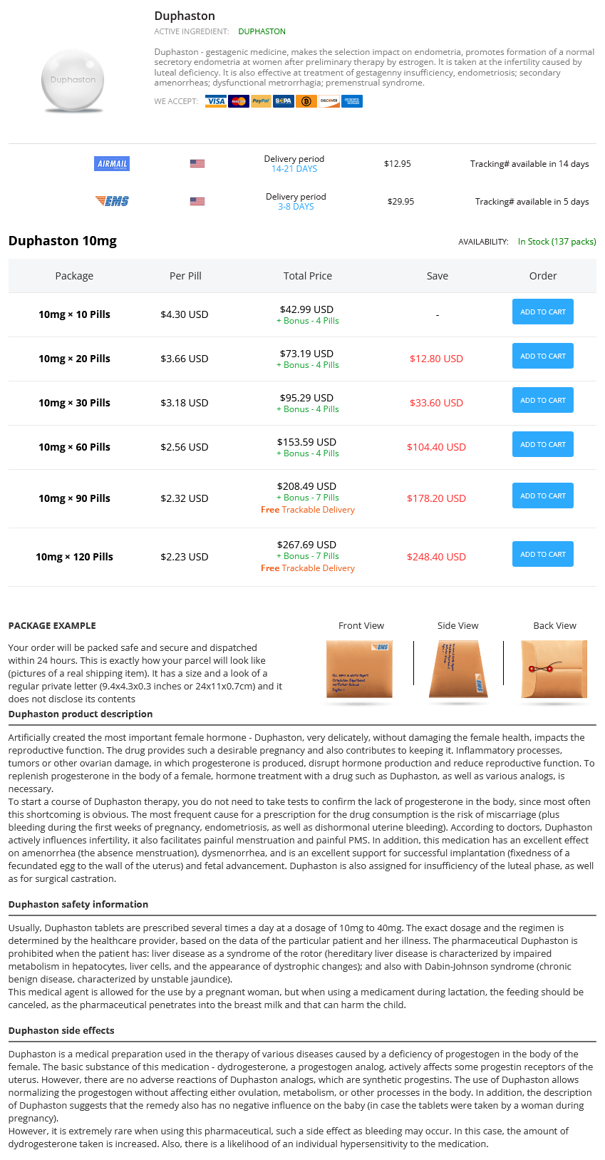
Duphaston Dosage and Price
Duphaston 10mg
- 10 pills - $42.99
- 20 pills - $73.19
- 30 pills - $95.29
- 60 pills - $153.59
- 90 pills - $208.49
- 120 pills - $267.69
Quantitative assessment of sensing and sequestration of spherocytic erythrocytes by the human spleen menstruation yeast infection purchase 10mg duphaston with amex. Macrophage plasticity and interaction with lymphocyte subsets: cancer as a paradigm. An unrestrained proinflammatory M1 macrophage population induced by iron impairs wound healing in humans and mice. Direct comparison of Sn-mesoporphyrin, an inhibitor of bilirubin production, and phototherapy in controlling hyperbilirubinemia in term and near-term newborns. Limited role for the bilirubin-biliverdin redox amplification cycle in the cellular antioxidant protection by biliverdin reductase. Dysfunction of the heme recycling system in heme oxygenase 1 deficient mice: effects on macrophage viability and tissue iron distribution. Membrane lipid composition and vesicle size modulate bilirubin intermembrane transfer. Regulation by heme of mitochondrial protein transport through a conserved amino acid motif. Characterization of recombinant human erythropoietin produced in Chinese hamster ovary cells. A common model for cytokine receptor activation: combined scissor-like rotation and self-rotation of receptor dimer induced by class I cytokine. Jak2 deficiency defines an essential developmental checkpoint in definitive hematopoiesis. The distal region and receptor tyrosines of the Epo receptor are non-essential for in vivo erythropoiesis. Involvement of phosphatases in proliferation, maturation, and hemoglobinization of developing erythroid cells. Mutation in murine erythropoietin receptor induces erythropoietin-independent erythroid proliferation in vitro, polycythemia in vivo. Apoptosis protection by the Epo target Bcl-X(L) allows factor-independent differentiation of primary erythroblasts. Effects of recombinant erythropoietin on murine megakaryocytic colony formation in vitro. Simultaneous measurement of reticulocyte and red blood cell indices in healthy subjects and patients with microcytic and macrocytic anemia. Identification of a functional role for lipid asymmetry in biological membranes: phosphatidylserine-skeletal protein interactions modulate membrane stability. Analysis of integral membrane protein contributions to the deformability and stability of the human erythrocyte membrane. Skubitz Specific (Secondary) Granules Although some specific (also called secondary) granules, like azurophilic granules, fuse with phagocytic vesicles, it is believed that these granules are largely for release into the extracellular space. For example, collagenase may degrade collagen, thus augmenting movement through collagen and participating in tissue remodeling. Apolactoferrin, by binding iron, may have an antibacterial effect by preventing bacteria from obtaining necessary iron for growth. When cells are stimulated, the surface expression of many of these membrane proteins is increased, and some of the upregulated molecules may be derived from specific granules. The importance of the specific granules in neutrophil function is shown in patients who lack specific granules; these patients are susceptible to repeated skin and respiratory infections and have defective neutrophil chemotaxis and adhesion. Many studies have been performed to identify and characterize the molecular composition of the various subcellular compartments of neutrophils. Two techniques are commonly used for this purpose: immunoelectron microscopy and subcellular fractionation. Most recent subcellular fractionation studies used the technique of nitrogen cavitation to disrupt the neutrophils. In this technique, cells are equilibrated with nitrogen at high pressure and then released into a pressure of 1 atm. The rapid decrease in extracellular pressure results in disruption of the cells by the formation of nitrogen gas within the cell. Although the granules of neutrophils may be best viewed as a continuous spectrum of granules, possibly resulting from differential production of different granule contents during neutrophil development, studies of neutrophils have operationally defined several types of granules. Four well-defined types of granules in neutrophils are primary granules, secondary granules, tertiary granules, and secretory vesicles, although additional heterogeneity of these fractions may exist. These granules fuse with phagocytic vesicles, resulting in the delivery of their contents to the ingested organism. Neutrophils are so named because of their neutral staining with Wright stain, whereas eosinophils avidly stain with the dye eosin, and basophils have readily identified large darkstaining granules with Wright stain. Neutrophils play a critical role in host defense by phagocytizing and digesting microorganisms, and inappropriate activation of neutrophils may result in damage to normal host tissues. In the resting uninfected host, the production and elimination of neutrophils are balanced, resulting in a fairly constant concentration of neutrophils in peripheral blood. When an infection occurs, chemotactic agents are generated that result in migration of neutrophils to the site of the infection and activation of neutrophil defensive functions. This effect is often associated with an increased production and release of neutrophils from the bone marrow. This chapter reviews the structure, morphology, production, distribution and kinetics, and functions of neutrophils.